Easily missed orthopaedic fractures in GP setting
Written 24th February 2020 - Last Update 29th July 2026
Easily missed fractures
See Fracture Immobilisation & Casting for general splinting/casting technique, review timing and referral thresholds — this page covers fracture-specific recognition and pitfalls.
Scaphoid fracture
Most often an axial loading injury to the thumb (FOOSH), but can follow a direct blow. Fractures are frequently undisplaced and invisible on initial X-ray — if clinically tender in the anatomical snuffbox or on axial thumb loading, treat as a scaphoid fracture regardless of X-ray: short arb backslab - Thumb spica no longer needed.
- Proximal-pole fractures carry high avascular necrosis risk — proximal 1/3 ≈ 33% AVN risk; proximal 1/5 (the pole itself) ≈ 100%.
- Management (immobilisation vs surgical fixation) is guided by stability, per orthopaedics; hand physiotherapy helps post-injury.
Radial head fracture
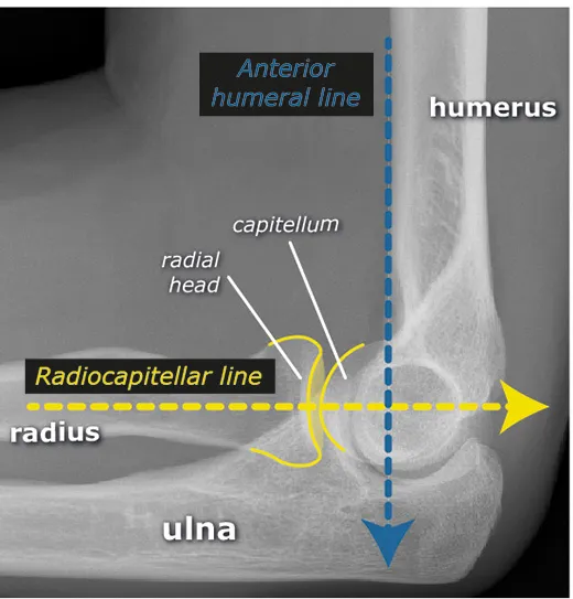
1–4% of upper limb fractures; FOOSH with axial load through the radial head. Easily missed without adequate views.
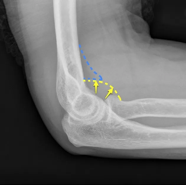
- Fat pad (“sail”) sign indicates a minimally displaced, otherwise occult fracture — the posterior fat pad is always abnormal if visible; the anterior fat pad is only abnormal if enlarged.
- Treatment is usually non-operative unless significantly comminuted or dislocated; long-arm backslab (above-elbow, bent) if immobilising.
Calcaneal and subtalar fractures
Axial load — falling onto straightened legs from height, or direct foot impact.
- Böhler’s angle (lateral view): line from the highest point of the anterior process to the highest point of the posterior facet, and a line tangential to the superior tuberosity edge. Normal 20–40°; a decreased angle indicates posterior-facet collapse.
- Angle of Gissane: line along the lateral margin of the posterior facet and a line anterior to the beak of the calcaneus. Normal 120–145°; an increased angle likewise reflects posterior-facet collapse.
- CT is the gold-standard imaging.
Lisfranc injury
A tarsometatarsal fracture-dislocation — traumatic disruption between the medial cuneiform and the base of the 2nd metatarsal. Mechanism: MVAs, falls from height, athletic injury via indirect rotational force + axial load through a hyperplantarflexed forefoot. Get a weight-bearing X-ray — missed injuries progress to chronic foot deformity and dysfunction, so maintain a high index of suspicion (see also foot fractures).
Five critical radiographic signs of midfoot instability (diagnostic on AP/oblique/lateral views): discontinuity of the line from the medial base of the 2nd MT to the medial middle cuneiform; widening of the 1st–2nd intermetatarsal interval ± a “fleck sign” avulsion fragment; dorsal displacement of the proximal 1st/2nd MT base; medial base of the 4th MT not aligning with the medial cuboid; disruption of the medial column line.
Supracondylar fracture (paediatric)
The most common paediatric traumatic fracture, age 5–7, equal sex incidence. 95–98% extension type, 5% flexion type; mechanism FOOSH (extension) or a fall on the olecranon (flexion). Often accompanied by an ipsilateral radius fracture. Minimally displaced fractures may show only a fat-pad sign on X-ray — immobilise in a backslab and refer (urgency depends on type).
Associated neuropraxia (nearly all resolve spontaneously after reduction/fixation):
- Anterior interosseous nerve (most common) — can’t make the “OK” sign
- Radial nerve (2nd most common) — sensory loss over the volar index finger
- Ulnar nerve (flexion-type injuries) — can’t extend wrist MCP or thumb IP joints
Vascular compromise occurs in 5–17% but is often mitigated by rich collateral circulation.

| Gartland type | Features | Management |
|---|---|---|
| I | Non-displaced. Beware subtle medial comminution → cubitus varus (technically not type I — needs reduction/pinning) | Cast immobilisation 3–4 weeks, X-ray at 1 week |
| II | Displaced, posterior cortex/periosteal hinge intact, sagittal-plane deformity only | Typically closed reduction + percutaneous pinning (CRPP) |
| III | Displaced, often 2–3 planes | CRPP, or open reduction if needed |
| IV | Complete periosteal disruption, unstable in flexion and extension (diagnosed under anaesthesia) | CRPP, or open reduction if needed |
| Medial comminution | Collapse of the medial column, loss of Baumann angle → varus malunion (“gunstock” deformity) | CRPP, often needs significant valgus force to reduce |
| Flexion type | Fall on the olecranon | CRPP, more likely to need open reduction |
Seymour fracture
A displaced Salter-Harris II fracture of a distal phalanx at the physis, from an axial stubbing injury forcing the digit into hyperflexion — in the lower limb this is usually the great toe. Easily missed because the fracture line and any germinal-matrix laceration are hidden under the nail; the only clue may be a small subungual/proximal nail-fold haematoma, pain, and (if the toe) a limp. Multi-view X-ray shows a Salter-Harris II fracture with flexion deformity at the fracture site.
This is functionally an open fracture — the nail bed/germinal matrix is typically interposed in the fracture gap, seeding it with skin flora — so it needs prompt irrigation, debridement of the interposed tissue, fracture reduction and antibiotics, not just a splint or buddy-strap. Refer rather than manage as a routine toe injury.
Related pages
- Basic fracture management (general principles) — see lower limb fractures, knee fractures & dislocations, shoulder injuries, forearm / elbow injuries, wrist / hand injuries, ankle fractures & dislocations, foot fractures
- Head, spine & chest fractures
- Stress fractures
- Approach to the Limping Child (Seymour fracture and plantar puncture wounds in the context of a paediatric limp)
- Fingertip & Nail Bed Injuries (finger Seymour fracture management, subungual haematoma, nail bed laceration)