Foot Fractures
Written 17th February 2020 - Last Update 29th July 2026
Foot fractures seen in urgent care/GP practice. For the Ottawa Foot Rules (when to X-ray at all), see Ankle Sprain, which covers both the ankle and foot criteria. ~53% of all fractures missed on plain films by a radiologist occur in the foot — X-rays of the foot and ankle are often ordered together because the two are hard to separate anatomically, so review both systematically on every significant foot/ankle injury.
Foot X-ray — what to check
- AP view: check the base of the 2nd metatarsal aligns with the 2nd cuneiform; assess the metatarsal/phalanx cortices medial to lateral for disruption (mnemonic “B5” — work proximal→distal assessing cortical surfaces (Bones), then check the base of the 5th metatarsal, the most commonly fractured bone in the foot, in detail).
- Lateral view: best view for the calcaneus and Böhler’s angle (below); also the best view to evaluate tarsometatarsal (Lisfranc) alignment.
- Oblique view: best for 5th metatarsal fractures; joint spaces around the cuboid should be equal and open.
- Weight-bearing views are increasingly important and can reveal instability (e.g. Lisfranc widening) that a non-weight-bearing film misses entirely — if plain films aren’t conclusive, don’t stop there.
Lisfranc injury
Tarsometatarsal fracture-dislocation — disruption of the joint between the midfoot and forefoot, classically at the 2nd metatarsal base/middle cuneiform (the “keystone” of the midfoot arch, stabilised by the Lisfranc ligament). A continuum from a subtle sprain to an obviously unstable fracture-dislocation.
- Mechanism: direct crush, or an axial load through a hyperplantarflexed or rotated foot (twisting on a planted foot, or a fall from height).
- ~20–25% are missed at first presentation — maintain a high index of suspicion, especially with midfoot swelling/pain out of proportion, pain with weight-bearing, and plantar bruising/ecchymosis under the midfoot (a specific sign).
- Alignment check on a normal AP foot X-ray: the lateral border of the 1st metatarsal should line up with the lateral border of the medial cuneiform; the medial border of the 2nd metatarsal should line up with the medial border of the middle cuneiform; the medial border of the 4th metatarsal should line up with the medial border of the cuboid. Any disruption raises suspicion for a Lisfranc injury. A “fleck sign” — a small avulsion fragment from either the medial cuneiform or the base of the 2nd metatarsal — is a subtle but important clue.
- A >2mm diastasis between the base of the 1st and 2nd metatarsals is equivalent to a 3rd-degree sprain/complete ligamentous rupture.
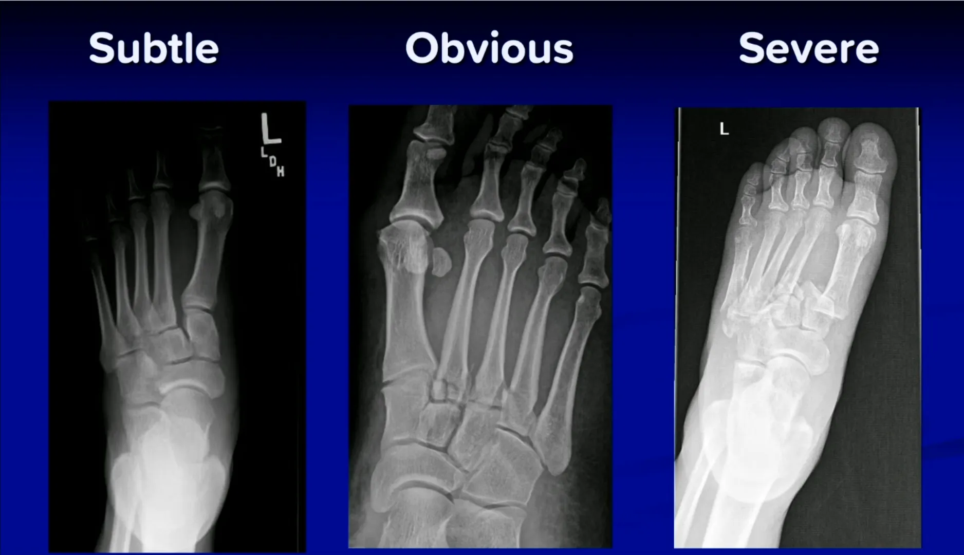
Imaging: if plain films are non-diagnostic but suspicion remains, get a weight-bearing view first (a standing/weight-bearing AP foot view can reveal widening a non-weight-bearing view misses entirely), then escalate to CT → MRI if still suspected. Once confirmed, refer promptly to orthopaedics for surgical treatment and place in a short-leg backslab (or a CAM boot) while awaiting definitive imaging/review.
Calcaneal fracture
Sustained from an axial load — landing on the feet after a fall from height, or in a motor vehicle collision (also consider a lumbar spine fracture — associated in 10–15% of cases — and always check the other foot, since 9% are bilateral). 75% are intra-articular (involving the posterior facet).
- Böhler’s angle — measured on the lateral view, normal 20–40°; a reduced angle indicates loss of calcaneal height and suggests a compression fracture.
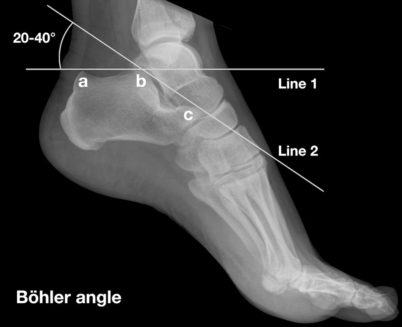
- Angle of Gissane (also lateral view) — between the lateral margin of the posterior facet and a line anterior to the beak of the calcaneus, normal 120–145°; an increased angle likewise reflects posterior-facet collapse (the opposite direction to Böhler’s angle) — the two are complementary, not interchangeable.
- Mondor sign — mid-plantar ecchymosis — may be present and is a useful clue to a subtle calcaneal fracture even when swelling is the only other finding.
- Tongue-type fractures (Essex-Lopresti pattern — the fracture fragment is displaced posterosuperiorly by the Achilles insertion) are tissue- threatening and an orthopaedic emergency — the displaced fragment can compromise the overlying skin, similar to a displaced ankle fracture with tenting.
- These are high-energy injuries — essentially all calcaneal fractures need ED/trauma evaluation and orthopaedic input, not routine GP follow-up; the role in urgent care is recognising the injury (Böhler’s angle, Mondor sign, mechanism) and arranging urgent transfer/imaging rather than definitive management.
Talar fracture
Uncommon but high-stakes — the talus has a precarious, largely retrograde blood supply, so displaced fractures carry a real avascular necrosis risk. A 1mm lateral shift of the talus can reduce joint contact area by as much as 42%, which is why even small-looking displacement matters.
- Talar neck fractures — graded by the Hawkins classification (based on displacement); risk of AVN rises steeply with each grade. All talar neck fractures displaced >2mm require operative repair.
- Talar head/body fractures are harder to see on plain films and often need CT.
- Any suspected talar fracture warrants orthopaedic referral given the AVN risk — this isn’t a “cast and review” injury.
Metatarsal fractures
Base of 5th metatarsal — three distinct zones, easily confused but managed very differently:
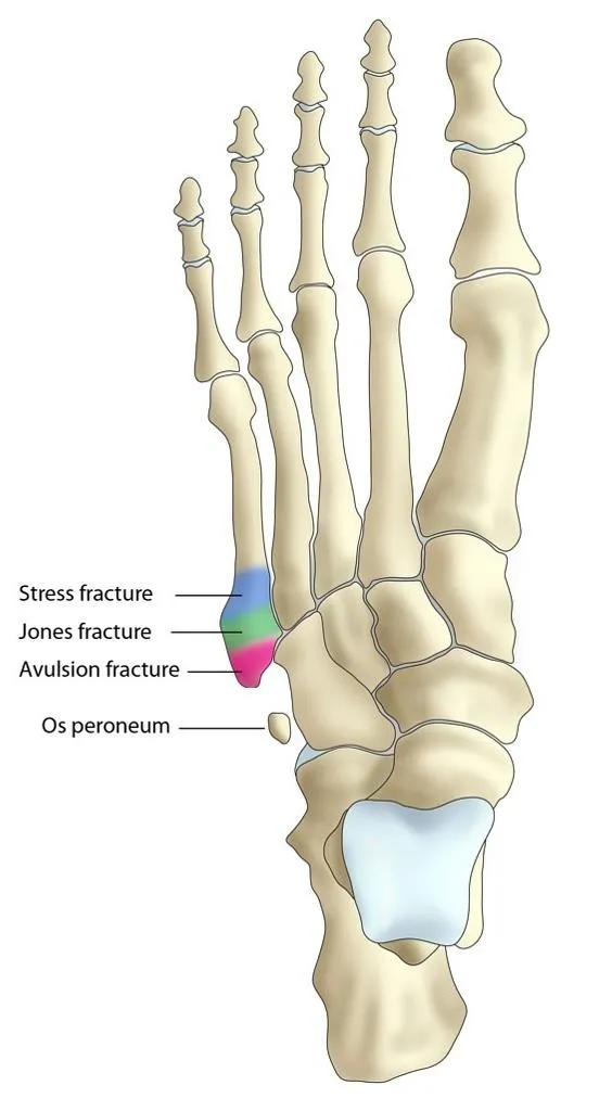
| Zone | Name | Mechanism/features | Management |
|---|---|---|---|
| Zone 1 | Avulsion fracture (“pseudo-Jones”/dancer’s fracture) | Avulsion of the peroneus brevis insertion at the tuberosity, from an inversion injury | Good blood supply, heals well — treat like an ankle sprain (boot, weight-bearing as tolerated) |
| Zone 2 | Jones fracture | Metaphyseal-diaphyseal junction — a watershed blood-supply zone | High non-union rate (30–50%); non-weight-bearing cast 6–8 weeks; often takes >2 months to heal; surgery favoured in active patients given the non-union risk |
| Zone 3 | Proximal diaphyseal stress fracture | Repetitive load, often in athletes | Non-weight-bearing (crutches/splint) — treatment (cast vs operative) depends on activity level |
Other metatarsal/phalangeal fractures: displacement in dorsal/medial/ lateral directions can be seen on both AP and lateral views. Stress fractures most often affect the 2nd/3rd metatarsals and are frequently occult on the initial film — if suspected clinically with a normal X-ray, treat empirically (stiff-soled shoe/boot, reduced activity) and reimage or escalate if symptoms persist.
Toe fractures
Buddy strapping for non-displaced fractures. Displaced intra-articular fractures — especially of the great toe — may need internal fixation.