Ankle Sprain
Written 22nd February 2020 - Last Update 5th September 2026
Ankle Sprain
(Consolidates the former Sprained Ankle” note.) The most common sporting injury (~25% of sports injuries). “Up to 90% involve the lateral ligaments — ATFL > CFL > syndesmosis > deltoid (the stronger, tauter medial deltoid ligament is less prone). High ankle sprain (syndesmosis injury) accounts for 1–10%. Ankle sprains are the most common reason for missed athletic participation. Limited dorsiflexion, decreased proprioception and balance deficiency all raise the risk of a fresh sprain.
Commonly associated fractures: base of 5th metatarsal, distal fibula, talus (including talar-dome chondral injury), calcaneus. Children <12 and the elderly are more likely to have fractured than sprained.
Mechanism and ligaments
Most sprains occur with the ankle plantar flexed and inverted — an awkward landing or uneven ground.
- ATFL — most commonly injured; plantar flexion + inversion; drawer laxity in plantar flexion.
- CFL — second; dorsiflexion + inversion; drawer laxity in dorsiflexion (contributes to subtalar stability).
- PTFL — rarely involved.
History
Ankle “gives way”; pain and difficulty weight-bearing; bruising (may take 12–24 hrs) indicates a more severe injury; recurrent sprains → functional instability on uneven ground. Pain decreases rapidly over the first 2 weeks, but 5–33% report some pain at 1 year, with increased risk of re-sprain on either side.
Examination
- Swelling/bruising — classically a rounded swelling in front of the lateral malleolus; palpate the three lateral ligaments and bony landmarks (Ottawa).
- Anterior drawer test (ATFL in plantar flexion, CFL in dorsiflexion) — limited usefulness acutely; talar tilt test (excess inversion vs the other side = ATFL+CFL).
- On the talar tilt test, more than 15° of excess inversion compared with the contralateral side indicates injury to both the ATFL and CFL.
- Syndesmosis squeeze test — squeeze fibula and tibia together at mid-calf: anterior ankle pain at the syndesmosis level.
- Too painful? Re-examine in 1–2 weeks.
Investigations — Ottawa Ankle Rules
X-ray only if pain in the malleolar zone plus bony tenderness at the posterior edge/tip of either malleolus (distal 6 cm) or inability to walk 4 steps; or pain in the midfoot zone plus tenderness at the navicular or 5th metatarsal base or inability to walk 4 steps. 96–99% sensitive for fracture. Be cautious with intoxication, distracting injuries, gross swelling, and age <18.
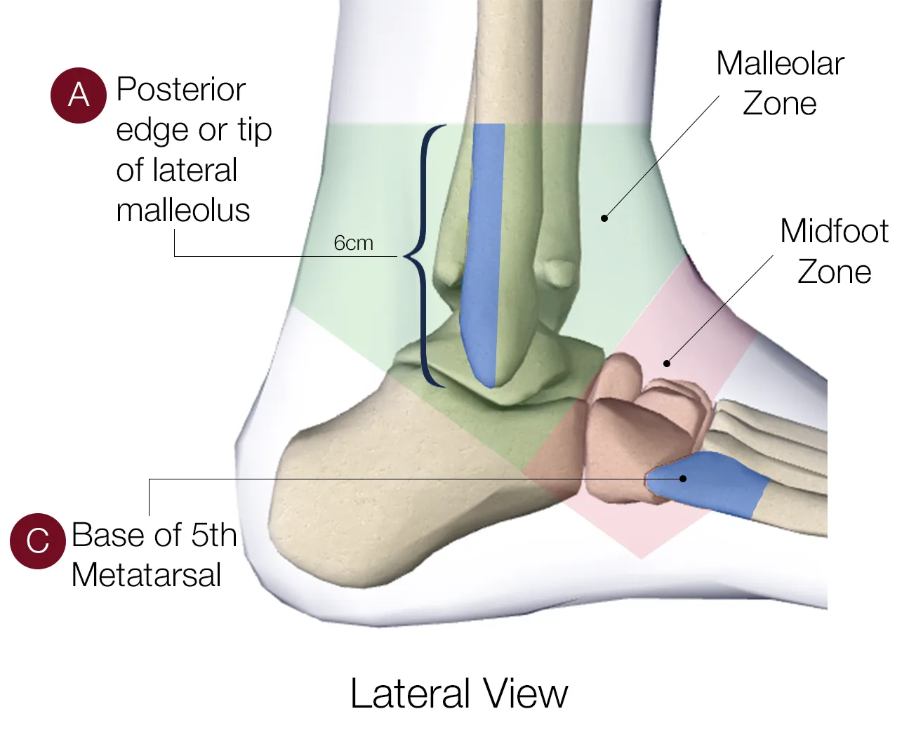
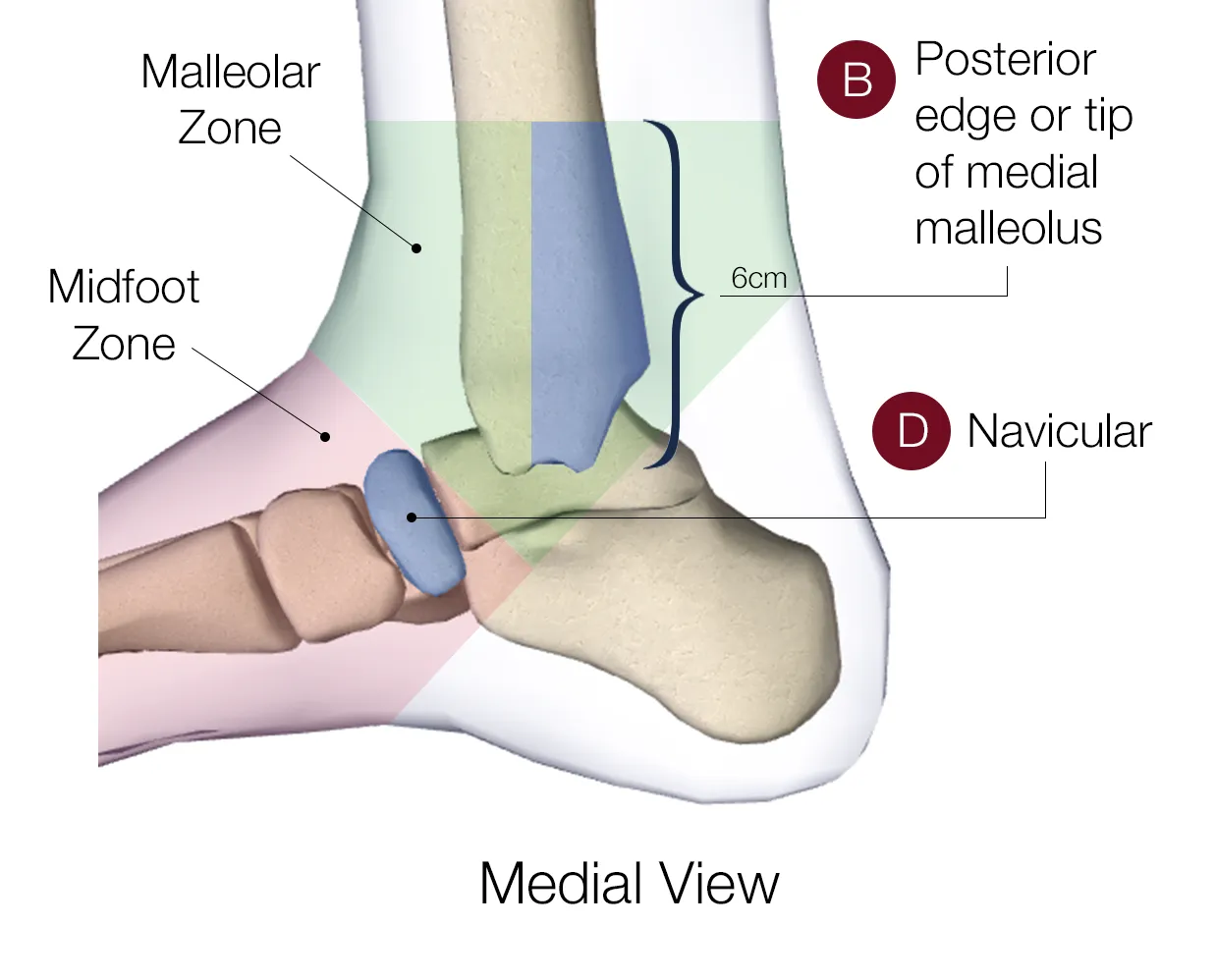
For the fracture patterns an ankle X-ray actually turns up — Weber classification, Maisonneuve, dislocation — see Ankle Fractures & Dislocations.
MRI if pain persists 6–8 weeks post-injury: peroneal tendon pathology, osteochondral injury, syndesmotic injury (X-ray cannot exclude syndesmosis).
Grading and management
For the general principles behind this table — protection vs prolonged rest, ice/compression evidence, and NSAID vs paracetamol vs opioid choice — see Acute Soft-Tissue Injury Management.
| Grade | Ligament | Findings | Management | Healing |
|---|---|---|---|---|
| I | Stretch, no tear | Minimal swelling, normal weight-bearing | Tubigrip/crepe, early mobilisation, review 1 week if not improving | 1–2 weeks |
| II | Partial tear | Moderate swelling/bruising, mild pain on weight-bearing | ± CAM boot 1 week, ± crutches 1–2 weeks, physiotherapy, review 1 week | 3–6 weeks |
| III | Complete tear | Severe swelling, severe pain/inability to weight-bear, gross instability on stress testing | CAM boot up to 4 weeks, ± crutches (WBAT), weekly review, physiotherapy ± taping | 6–12 weeks, possibly longer |
Most acute ankle sprains, including virtually all grade I and II and most grade III sprains, are managed nonoperatively, with the goal of allowing ligament healing while maintaining range of motion, strength and stability. A short period of immobilisation (e.g. cast or boot, non-weight-bearing) can help a grade III sprain too painful to walk on, but extended immobilisation beyond that initial period is discouraged — casting for more than 4 weeks is not recommended, even for the most severe sprains, because prolonged immobilisation can cause weakness and stiffness without improving long-term outcomes. A semi-rigid brace, air stirrup, or taping is preferred once safe, both to protect the ligament and to restore proprioceptive feedback; functional bracing or taping for 4–6 weeks after a sprain is more effective than prolonged casting at preventing recurrent injury.
- RICE + NSAID (e.g. ibuprofen 400 mg tds with food; 10 mg/kg in children), then graded return to activity — early functional rehabilitation beats prolonged immobilisation.
- Syndesmosis injury → acute orthopaedic assessment.
- Non-acute orthopaedic referral: uncertain diagnosis, no improvement at 3–6 months, or instability unresponsive to physiotherapy (options include anatomic reconstruction; arthroscopic debridement for impingement lesions).
Therapeutic ultrasound has no proven benefit. A Cochrane review of 6 trials (606 participants) found ultrasound therapy does not enhance recovery, reduce pain and swelling, or improve the ability to stand on the affected foot after an ankle sprain, and any residual benefit is probably too small to be clinically important.
Chronic lateral ankle instability
Chronic lateral ankle instability occurs in 10–20% of people after an acute ankle sprain. Initial treatment remains conservative — bracing or a structured neuromuscular training programme, which gives short-term improvement in functional stability, though longer-term follow-up data are limited. If conservative management fails and ligament laxity persists, surgery is considered: the gold-standard is an anatomic repair of the torn ATFL ± CFL (Broström repair, often reinforced with the Gould modification), with good-to-excellent outcomes in ~90% of patients and most athletes returning to play by 3–4 months. Evidence does not clearly favour one surgical technique over another, but early functional rehabilitation after surgical reconstruction leads to an earlier return to work and sport than six weeks of immobilisation.
Prognosis and complications
While most people recover fully, up to 40% of patients experience residual symptoms, including chronic instability from insufficient ligament healing or proprioceptive deficits, recurrent sprains, or (less commonly) post-traumatic degenerative joint changes. Consistent early rehabilitation (balance and strength training) reduces the risk of reinjury. Prolonged immobility or swelling after a severe sprain can also lead to blood clots — a further reason to favour early functional treatment over extended casting.