Ankle Fractures & Dislocations
Written 17th February 2020 - Last Update 27th August 2026
This page covers Ankle fractures and dislocations, for ligamentous ankle sprain (including the Ottawa Ankle Rules and syndesmosis sprain grading), see Ankle Sprain
Ankle fractures range from minor avulsions to large trimalleolar fractures from high-velocity trauma. A fracture involving just 1 malleolus tends to be stable; 2 or more fractured malleoli are considered unstable. Always examine the entire tibia/fibula (palpate proximally, especially the fibular head/neck) and the knee, and have a high suspicion for an associated syndesmotic injury with any ankle fracture — missed fractures/instability lead to chronic ankle instability, loss of ROM, and post-traumatic arthritis.
Ankle X-ray Interpretation
- AP view: mortise should be an equal width all the way round (unequal → look for a fracture); tibiofibular overlap should be >6mm (or clear space <6mm) — less overlap than this suggests a syndesmosis injury/ligamentous disruption/fracture somewhere. Mnemonic: “M&M overlap” — malleoli, mortise, overlap.
- Mortise view (foot internally rotated ~15–20°): medial clear space should be <4mm — widening suggests a deltoid ligament injury and an unstable joint likely needing surgical fixation. Also check the talar dome outline (irregularity → talar dome fracture) and the base of the 5th metatarsal.
- Lateral view: look for posterior malleolus fractures (can be subtle), check the tibiotalar joint space, and — if a calcaneal fracture is suspected — Böhler’s angle (see Foot Fractures).
- Squeeze test (compress the mid-tibia and fibula together) for syndesmotic injury — do this on every ankle injury, not just Ottawa Ankle Rules.
- Isolated ankle dislocation without a fracture is uncommon — assess stability carefully when there’s clinical doubt.
Weber classification (lateral malleolus fracture)
Classifies fractures by their level relative to the ankle joint/syndesmosis — the syndesmosis is the key structure at risk with each higher grade.
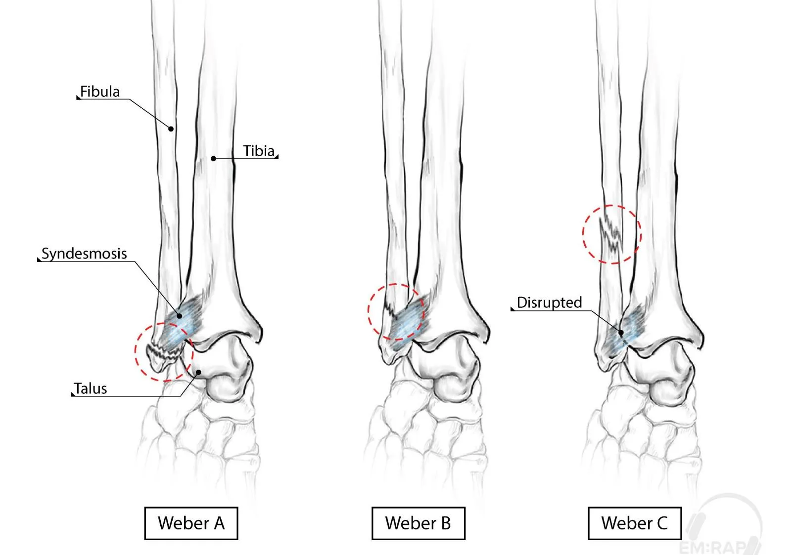
| Type | Level | Stability | Management |
|---|---|---|---|
| A | Below the ankle joint (syndesmosis intact) | Usually stable | Isolated, non-displaced distal avulsion fractures can be treated like an ankle sprain — boot, weight-bearing as tolerated. Ortho follow-up 1–2 weeks. |
| B | At the level of the joint | Unstable in up to 50% (depends on deltoid ligament integrity) — stress views may be needed | Posterior long-leg/backslab, non-weight-bearing, urgent ortho for operative consideration |
| C | Above the joint (syndesmosis disrupted) | Unstable | Posterior splint, non-weight-bearing, urgent ortho — usually needs ORIF |
Isolated, non-displaced medial malleolus fractures not involving the joint can also be treated non-operatively in a posterior splint, non-weight-bearing. Bimalleolar and trimalleolar fractures are inherently unstable and generally need open reduction and internal fixation.
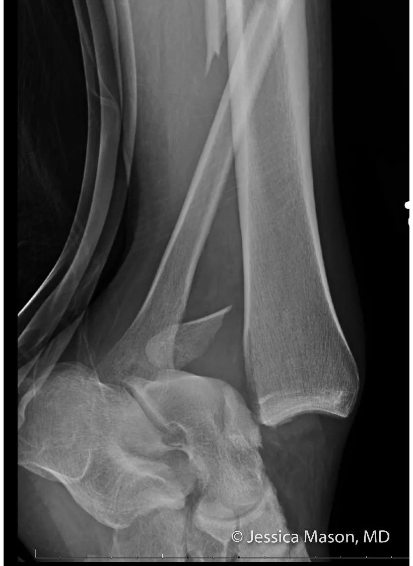
Weight-bearing views matter: a fracture that looks stable on a non-weight-bearing film can look unstable on a proper weight-bearing film — if there’s any doubt about stability, a weight-bearing view can change management.
Maisonneuve fracture
Medial malleolus fracture (or deltoid ligament rupture) + a fracture of the proximal fibula, from an external rotation force through the ankle that traverses the interosseous membrane and exits through the relatively weak superior fibula. Easily missed — patients often don’t volunteer pain at the knee/proximal leg, so always palpate the full length of the fibula, especially the fibular head, with any ankle injury (this isn’t part of the Ottawa Ankle Rules).
- Associated with peroneal nerve injury (the common peroneal nerve winds around the fibular neck) — foot drop and loss of sensation in the first dorsal web space (between the 1st/2nd toes).
- If there’s tenderness over the medial malleolus, or a medial malleolus fracture, X-ray the entire leg including the knee. If there’s proximal leg tenderness or medial clear space widening without an obvious ankle fracture, X-ray the tibia/fibula to rule out a Maisonneuve injury.
- Usually needs operative repair (syndesmotic screws). The fibular head/neck fracture itself typically doesn’t need fixation.
Ankle dislocation
Nearly always occurs with a fracture (isolated dislocation without fracture is uncommon and needs a large amount of force) — from a high-speed mechanism (MVA, fall, other high-energy trauma). Assess for neurovascular compromise (weak/absent dorsalis pedis or tibialis posterior pulses, poor capillary refill) and skin tenting (impending open injury/necrosis risk) — don’t delay reduction to get imaging if there’s neurovascular compromise or tenting.
Reduction (procedural sedation/analgesia first):
- Flex the hip and knee to 90° (relaxes the gastrocnemius/Achilles).
- Apply constant longitudinal traction to the foot, with an assistant applying countertraction on the thigh — anterior or posterior force as needed to reverse the direction of displacement.
- Recheck neurovascular status immediately after reduction.
- Splint in a 3-sided short-leg splint, elevate the leg, non-weight bearing.
Nearly all ankle dislocations need surgical stabilisation. If adequate reduction and mortise alignment are achieved and there’s no neurovascular compromise, the patient can usually be discharged in a splint with close ortho follow-up (ideally within a week) rather than admitted. Superior dislocations typically result in a pilon fracture (comminuted tibial plafond fracture — high-energy axial load, always needs orthopaedic input); in posterolateral dislocations the tibialis posterior tendon can block reduction and may need to be manually freed first.
Paediatric ankle fractures
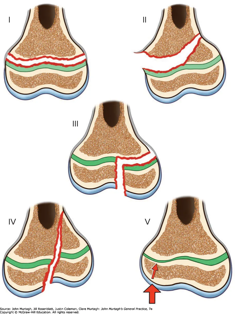
Growth-plate (physeal) fractures follow the Salter-Harris classification — higher grades carry more risk of growth arrest and generally need more aggressive management.
X-ray-negative inversion ankle injury in a child is much more often a sprain than a Salter-Harris I fracture of the distal fibula — older teaching (that children’s ligaments are stronger than their growth plates, so a fracture is more likely) has been superseded by newer data showing sprains are the more common injury. In practice this doesn’t change what you do: both a sprain and a non-displaced Salter-Harris I fracture are managed the same way — a removable brace, weight-bearing as tolerated, and follow-up — since advanced imaging to formally distinguish them isn’t necessary and removable bracing outperforms rigid splinting on functional recovery either way. A genuine high ankle sprain (syndesmosis injury — pain just above the ankle joint, worse on the squeeze test) needs longer immobilisation (6–8 weeks) and orthopaedic follow-up, unlike a routine lateral ligament sprain.
Tillaux fracture — a Salter-Harris III fracture of the antero-lateral distal tibial epiphysis, from forced external rotation. Occurs in the narrow window (12–14 years) where the distal tibial physis has already fused centrally and posteromedially but the antero-lateral portion is still open and vulnerable — closure of this physis proceeds centrally → anteromedially → posteromedially, sealing laterally last. Presents like a severe ankle sprain (anterolateral swelling/tenderness, pain on dorsi/plantarflexion, can’t weight-bear) but needs immediate orthopaedic referral — closed reduction is often attempted, and >2.5 mm of residual displacement generally needs surgical fixation.
Triplane fracture — a Salter-Harris IV injury of the distal tibia combining a vertical fracture through the epiphysis, a horizontal fracture through the physis, and an oblique fracture through the metaphysis (looks like a Salter-Harris II on the lateral view and a Salter-Harris III on the AP view — the two views need to be read together). Similar mechanism and age range to Tillaux; same immediate-referral principle. Both fractures need CT for pre-operative planning, and immediate orthopaedic referral for a neurovascular exam and reduction — don’t manage either as a sprain even though the initial presentation looks similar.