Fingertip & Nail Bed Injuries
Last Update 5th September 2026
Fingertip & Nail Bed Injuries
Crush, laceration and avulsion injuries to the fingertip are among the most common hand injuries seen in general practice and urgent care. Most are managed definitively without a hand surgeon.
For paronychia and other nail infections see Paronychia; for digital block technique see Local Anaesthesia.
Assessment
Ask the mechanism, hand dominance and occupation. Check tetanus status.
Examine sensation, capillary refill and active range of motion at the interphalangeal joints before injecting local anaesthetic. Look specifically for nail fold disruption, nail bed exposure and bone exposure. Three-view X-rays of the digit are indicated for any crush or laceration mechanism, since tuft and other distal phalanx fractures are commonly associated.
Subungual haematoma
A subungual haematoma follows a crush injury to the nail bed and presents with throbbing pain and blood visible under the nail.
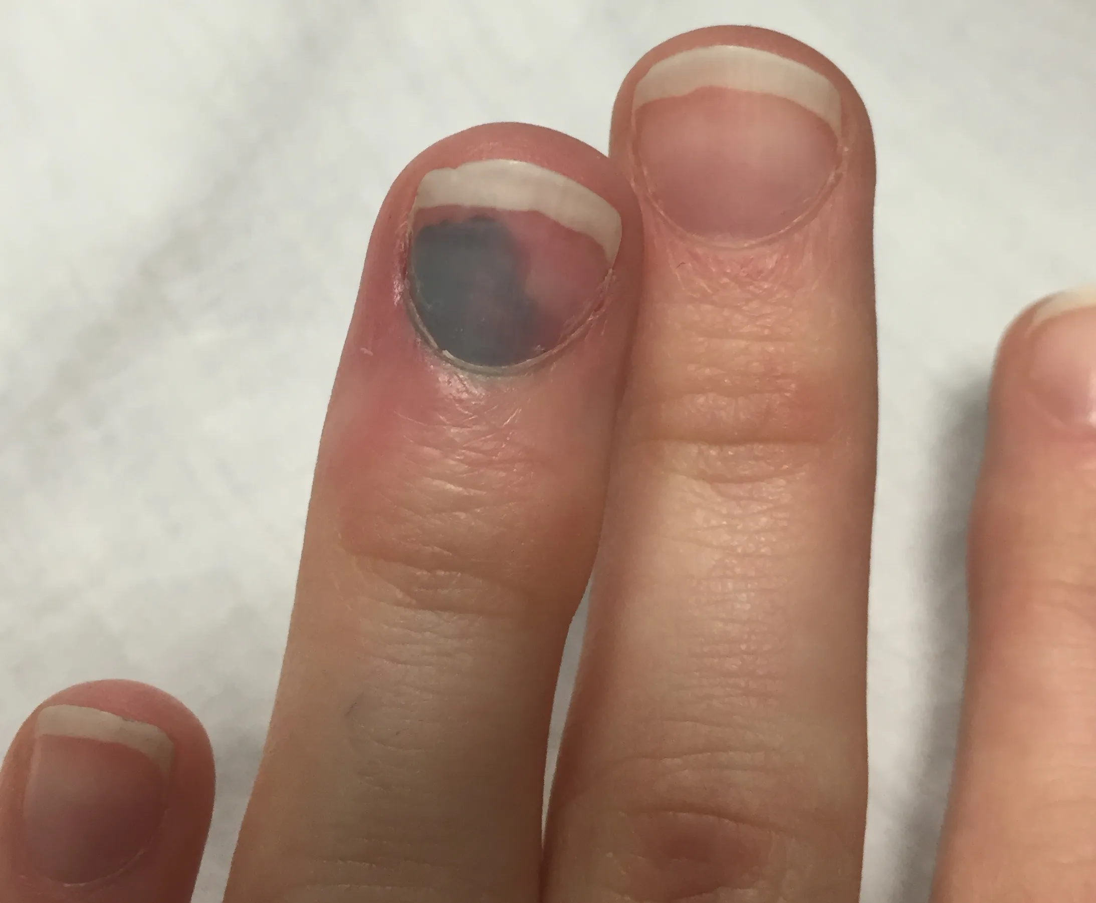
Image credit: Subungal hematoma of the finger.jpg — Callaleo, CC BY-SA 4.0, via Wikimedia Commons (https://commons.wikimedia.org/wiki/File:Subungal_hematoma_of_the_finger.jpg).

Trephinate within 48 hours of injury. Clean the nail with antiseptic; a digital block is usually unnecessary for trephination itself, since the procedure is typically less painful than the injection would be. Create a hole through the nail plate over the centre of the haematoma with electrocautery, an 18-gauge needle rotated in a drilling motion, or a heated paper clip, then let the trapped blood drain. Warn the patient that oozing for a day or two is expected, and that the haematoma will migrate distally with the nail over the following weeks — a haematoma that stops advancing needs review to exclude a pigmented lesion underneath.
Electrocautery is contraindicated over acrylic nails because of ignition risk. Routine post-procedure antibiotics are not supported by the evidence.
Nail bed laceration
Suspect an underlying nail bed laceration when the haematoma involves the whole nail, the nail itself is cracked, or the nail fold is disrupted — up to half of nail bed injuries have an associated distal phalanx fracture, so X-ray first.
Remove the nail plate to expose and repair the laceration with fine absorbable sutures (6-0 or 7-0 chromic gut), under a digital block. An RCT has shown tissue glue (2-octylcyanoacrylate) gives cosmetic and functional results comparable to suturing for nail bed repair, with a quicker repair time, and is a reasonable alternative in the appropriately trained operator’s hands. Trim the removed nail plate and reposition it under the nail fold as a stent, or use a non-adherent dressing instead, to keep the fold open for the new nail to grow through. Splint any associated fracture in extension for about four weeks with hand-therapy or hand-surgery follow-up.
Caution: trephinating a haematoma through an undisplaced distal phalanx fracture converts it to an open fracture, which can change management — check the X-ray before draining.
Tuft and distal phalanx fractures
Distal phalanx (tuft) fractures are usually stable and managed with an L-shaped or aluminium foam splint protecting the fingertip for two to three weeks. Refer for large soft-tissue loss or intra-articular involvement over 30% of the joint surface, and treat any associated nail bed injury as above.
Seymour fracture — don’t miss it in a child
A Seymour fracture is an open physeal (growth plate) fracture of the distal phalanx in a child, usually from a crush or hyperflexion mechanism at the nail fold. It can look like a simple mallet finger, but the deformity comes from displacement through the fracture rather than the DIP joint, and the nail fold laceration means the fracture is open — untreated it carries a high risk of infection and growth arrest. Any child with a distal phalanx fracture and a nail fold injury needs the nail lifted or removed to inspect for interposed nail bed or germinal matrix tissue, irrigation, antibiotics and same-day hand-surgery referral.
Amputation
Non-operative management (healing by secondary intention or simple primary closure) suits a wound with no exposed bone or tendon and less than about 2 cm of soft-tissue loss. Larger defects, or exposed bone that cannot be trimmed back without compromising nail bed support, need flap reconstruction and hand-surgery referral. Preserve any amputated tissue in saline-soaked gauze and transport it with the patient — even when replantation is not planned, it can sometimes still be used as a composite graft.
Refer urgently (same day) for: significant bone exposure, extensive laceration involving the eponychium, tendon or nerve injury, an open dislocation, or any amputation being considered for replantation.
High-pressure injection injury — do not underestimate
A puncture wound from a grease gun, paint sprayer or pressure washer looks trivial but is a hand-surgery emergency. Injected material tracks along tendon sheaths and fascial planes far beyond the visible entry point, and overall amputation rates after high-pressure injection injury run around 30–40%, higher again with organic solvents such as paint or turpentine and with delayed debridement.
Do not be reassured by a benign-looking puncture wound — diagnosis is often delayed while neurovascular compromise, oedema and compartment syndrome develop silently over the following hours. Give a tetanus booster and start broad-spectrum antibiotics; elevate the limb and leave the wound open, without a compressive dressing. Do not attempt a digital block, apply ice, or close the wound — all can worsen tissue ischaemia or mask deterioration. Arrange emergency transfer to a centre with hand-surgery theatre availability; early surgical debridement is the strongest predictor of avoiding amputation.
Follow-up and safety-netting
Warn every patient with a fingertip injury that swelling, cold intolerance and altered sensation can persist for months. Review nail bed repairs and splinted fractures at one to two weeks, and arrange hand-therapy referral for anyone with a repaired laceration, a fracture needing splint supervision, or stiffness developing during immobilisation.