Shoulder Fractures and Dislocations
Written 17th February 2020 - Last Update 6th August 2026
Traumatic shoulder injuries seen in urgent care/GP practice — clavicle, AC joint, proximal humerus, and scapula. For rotator cuff tears and impingement, see Rotator Cuff Tendinopathy; for glenohumeral dislocation, see Shoulder Dislocations / Stabilisation.
Always examine the joint above and below the injury, document neurovascular status before and after any splinting, and have a low threshold for a full 3-view series (AP, scapular Y, axillary) — a dedicated clavicle/scapula view if either is specifically suspected. Urgent referral for any open fracture, skin tenting (impending open fracture), or signs of neurovascular compromise.
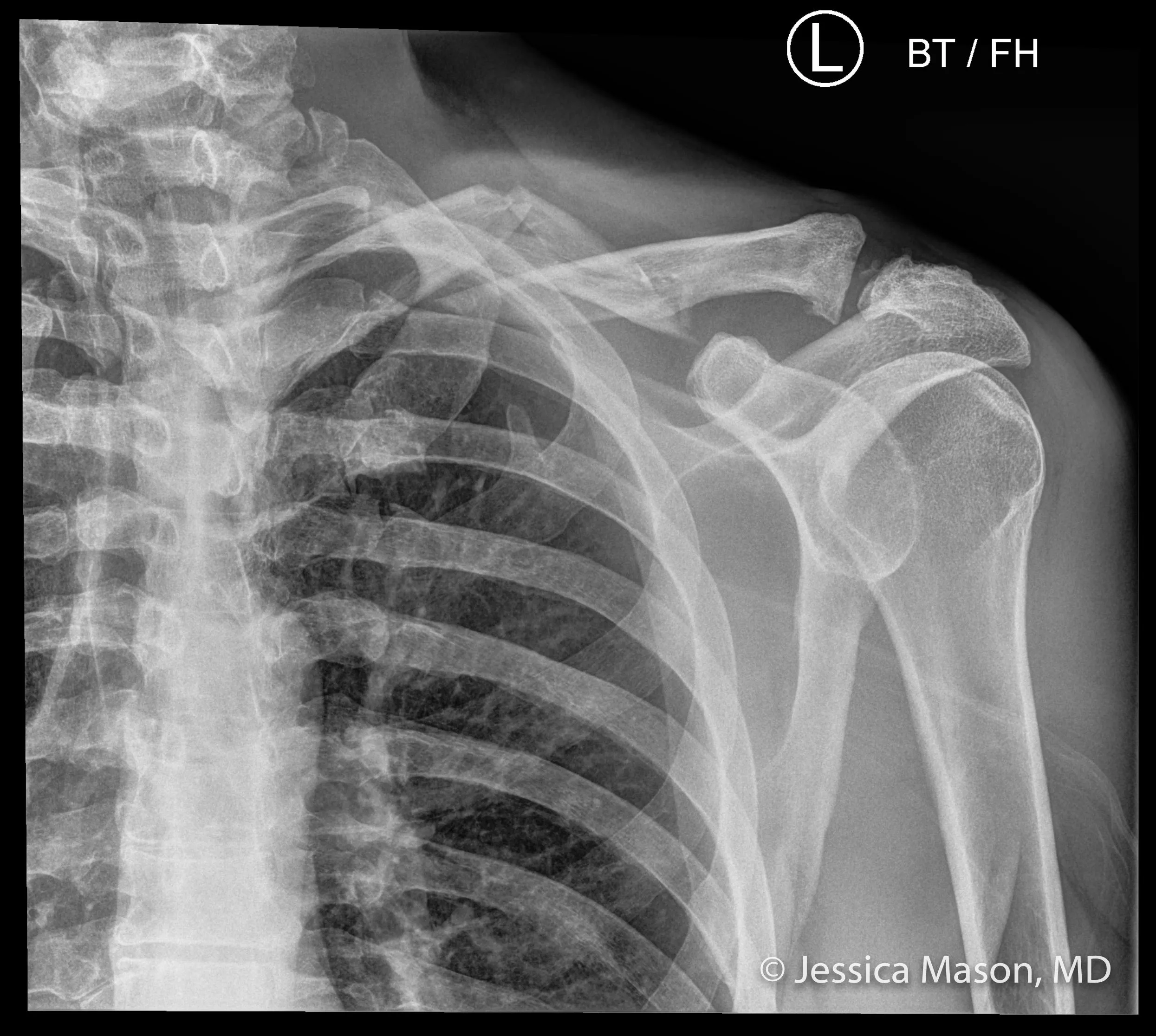
Clavicle fracture
The most common shoulder-girdle fracture — young males after a fall onto the shoulder/outstretched hand or direct blow, comprising 44–66% of shoulder injuries. Middle third (Allman type 1) accounts for 69–80%; lateral third (type 2) 21–28%; medial third (type 3) only ~2% but carries the highest risk of underlying vascular/lung injury (subclavian vessels, lung apex) — assess lung fields and distal pulses.
Management:
- Most are treated non-operatively in a simple sling (figure-of-eight bracing is no longer preferred — no better union rate, less comfortable).
- Refer for consideration of surgery if: displaced >100% (no cortical contact), >1.5cm shortening, comminuted, or involving the AC joint ligaments.
- Urgent referral if skin tenting is present (impending open fracture).
- Otherwise: sling, analgesia, ortho follow-up within ~1 week. Gentle ROM at 2–4 weeks, strengthening 6–10 weeks, return to sport 8–12 weeks — progress gradually to avoid adhesive capsulitis.
Acromioclavicular (AC) joint injury
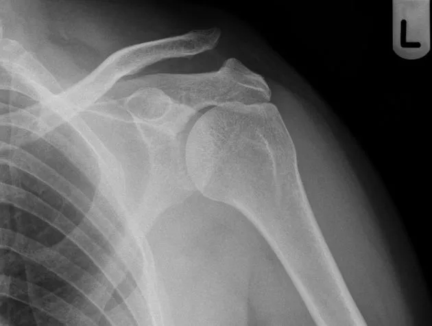
Direct blow to the point of the shoulder (fall with the arm adducted) or a FOOSH. Tenderness over the AC joint is the most reliable sign — Type I injuries often have a normal X-ray, so a good exam matters as much as imaging.
Exam: cross-body (horizontal) adduction test and O’Brien’s test — both can also be positive with AC joint OA, so interpret alongside the history.
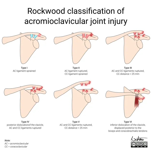
Rockwood classification (graded on X-ray by coracoclavicular (CC) and AC-joint distance, compared with the uninjured side):
| Type | Injury | Management |
|---|---|---|
| I | AC ligament sprain, normal X-ray | Sling, analgesia, physio for ROM/strength |
| II | AC ligament ruptured, CC ligament sprained | Sling, physio |
| III | Both AC and CC ligaments ruptured, CC distance <2x normal | Usually non-operative (controversial — surgery if high-demand/cosmetic concern); sling + physio, ortho follow-up |
| IV–VI | Posterior/inferior clavicle displacement or CC distance >2x normal | Refer for surgery — sling in the interim |
Proximal humerus fracture
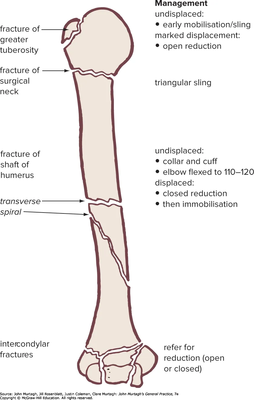
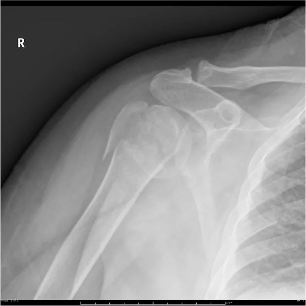
Typically an elderly fall onto an outstretched arm (2:1 female, osteoporosis/ epilepsy/diabetes as risk factors); higher-energy trauma in younger patients.
- Test axillary nerve function (deltoid abduction + “regimental badge” lateral-shoulder sensation) — the nerve most often injured, especially with subcoracoid dislocation of the humeral head.
- Risk of concurrent rotator cuff tear if the fracture involves the greater/lesser tuberosity insertion sites.
- Anatomical neck fractures carry a real avascular necrosis risk (the posterior humeral circumflex artery, the main blood supply to the head, is most vulnerable here).
- ~80–85% are managed non-operatively: sling immobilisation, early pendulum exercises within 2–3 weeks to prevent adhesive capsulitis, then progressive physio.
- Refer urgently for: 4-part fracture, anatomical neck fracture, or fracture-dislocation (Neer classification — displacement >1cm or angulation >45° between fragments defines a ‘part’).
Scapular fracture
Uncommon (~0.5% of all fractures) — the scapula is well protected by muscle, so a scapular fracture implies a significant-energy mechanism; assume concomitant polytrauma, especially chest (rib fractures ~64% co-occur) and spine, until proven otherwise. Most are managed non-operatively (sling, analgesia, physio) — surgery is rare (<1%), reserved for a “floating shoulder” (concomitant clavicle + scapular neck fracture), significant glenohumeral instability, or a displaced glenoid/neck fracture.
Shoulder dislocation (brief)
Anterior dislocation accounts for ~97% of shoulder dislocations; posterior is rare and easily missed on a single AP view (obtain a scapular Y or axillary view — look for loss of external rotation). Full assessment, reduction principles, and post-reduction rehabilitation are covered on Shoulder Dislocations / Stabilisation. A few points worth flagging for urgent care specifically: arterial injury is rare (<1%, most often the axillary artery) but should be reassessed for post-reduction; a neurological deficit (usually neurapraxia, with excellent recovery) is found in ~13.5% of patients after reduction; and traumatic shoulder dislocation is rare in children (<2%) before the humeral physis closes (14–17yo) — a skeletally immature patient with this mechanism is more likely to have sustained a proximal humerus fracture instead.