Lower Limb Fractures
Written 17th February 2020 - Last Update 13th August 2026
Lower Limb Fractures
Clinical decision rules and fracture patterns for the knee/lower leg. For knee and proximal-tibia fractures (patella, tibial plateau, tibial spine/ tuberosity avulsion) and true knee dislocation, see Knee Fractures & Dislocations. For ankle and foot fractures — Weber classification, Maisonneuve, ankle dislocation, calcaneal/talar/Lisfranc/metatarsal fractures, and the Ottawa Ankle/Foot Rules — see Ankle Fractures & Dislocations and Foot Fractures.
Ottawa Knee Rule
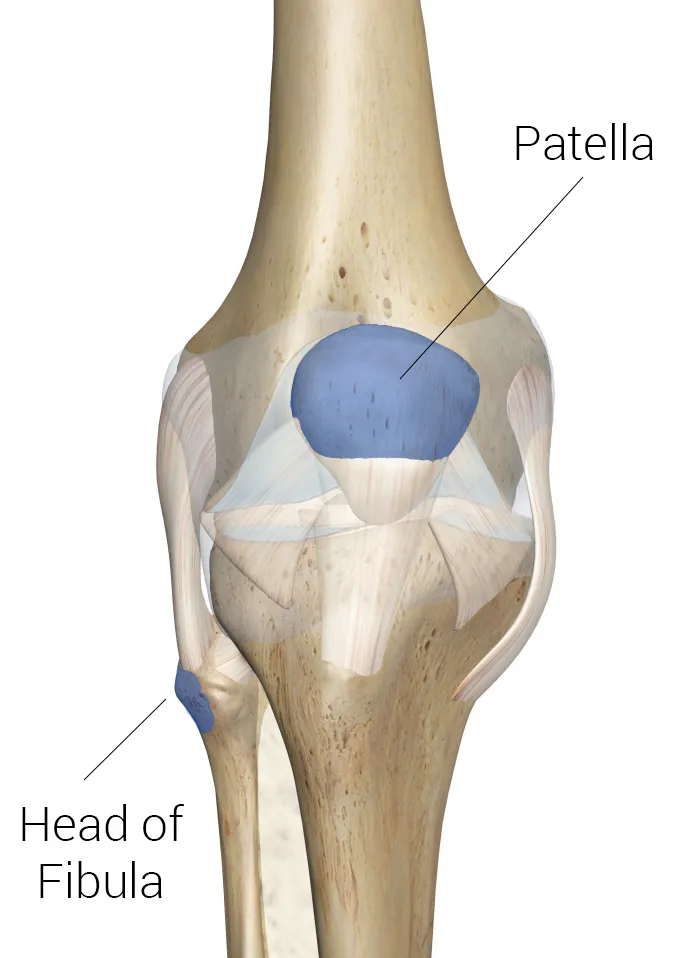
X-ray the knee if any of: age >55; isolated patellar tenderness (not significant with other tenderness present); fibular head tenderness; cannot flex to 90°; unable to weight-bear.
Tibial and fibular shaft fractures
Tibial shaft fractures carry the highest risk of compartment syndrome of any fracture — symptoms can take hours to develop, so any tibial shaft fracture needs serial reassessment, not a single check at presentation. For recognition and management of compartment syndrome itself, see Acute Compartment Syndrome. Because the tibia sits directly under the skin along most of its length, any skin break near or over the fracture should be treated as an open fracture (immediate antibiotics + urgent orthopaedic referral) even if it looks like an incidental puncture rather than an obvious compound injury.
- Mechanism: tibial shaft — high-velocity trauma in younger patients, or a simple low-velocity twisting injury in older patients; fibular shaft — classically a direct lateral blow. A history of a rapid increase in training load ± caloric restriction points to a stress fracture instead (X-ray is often initially normal — treat presumptively if the history/exam fit).
- Examination: point tenderness, deformity, swelling; always examine the knee and ankle for a concomitant injury. Full neurovascular exam — dorsiflexion/plantarflexion, toe extension/flexion, sensation over the anterolateral leg/dorsal foot/first web space (peroneal nerve — especially vulnerable where it wraps the fibular neck). For any ankle-pain-predominant presentation with a proximal fibular fracture, examine for a Maisonneuve fracture. Assess for syndesmosis injury: palpate for syndesmosis tenderness, squeeze test (compress tibia+fibula at mid-calf — pain at the ankle is a fairly specific but not very sensitive positive), and dorsiflexion-external rotation stress test (more sensitive, less specific) — no single test is enough alone, but the combination is useful, and a strong clinical suspicion warrants presumptive non-weight-bearing splinting even with all tests negative.
- Investigations: AP + lateral X-ray of the tibia/fibula, plus the knee and ankle if there’s any suggestion of a concomitant injury.
- Management:
- isolated, closed, non-displaced fibular shaft fracture — posterior short-leg splint, non-weight-bearing, outpatient orthopaedic follow-up; almost all can be managed this way.
- tibial shaft fracture (any displacement, or both-bone tibia/fibula) — long-leg posterior splint, strict non-weight-bearing, elevation, and urgent (same-day) orthopaedic referral — most need admission for monitoring and are high-risk for compartment syndrome regardless of how the fracture looks on X-ray.
- refer/transfer urgently for any open fracture, neurovascular compromise, or signs of current/impending compartment syndrome (increasing pain out of proportion, pain on passive toe stretch, increasing analgesia requirement). For open-fracture antibiotic timing, Gustilo grading and wound handling, see Open Fractures & Contaminated Limb Wounds.
- lower-leg immobilisation itself raises DVT risk — mention this to the patient and advise return if they develop calf swelling/pain or breathlessness; routine prophylaxis after immobilisation isn’t currently recommended, but discuss it for patients with other VTE risk factors.
Toddler’s fracture
A hairline spiral/oblique tibial fracture in a young ambulatory child (typically 9 months–3 years), from a low-energy twisting injury — often minimal or no witnessed trauma (a leg caught while running, or a jump with rotation). Presents with a limp or refusal to weight-bear; examination is often unremarkable (no swelling, no deformity), with only point tenderness over the tibial shaft.
Very commonly occult on the first X-ray — in one cohort, 39% of clinically-diagnosed toddler’s fractures had a negative initial film, and 93% of those later showed periosteal healing on a follow-up film. An internal oblique view picks up some fractures missed on standard AP/lateral views. If imaging is negative but the history/exam fit, splint for comfort and either repeat the X-ray in ~10 days or simply treat presumptively.
Management is shifting away from rigid casting — a walking boot for 3 weeks gives similar outcomes to a backslab/cast for non-displaced fractures, without needing orthopaedic follow-up, and with a faster return to activity — though one small study found a trend toward more early pressure-related skin complications with the boot than with casting, so this isn’t a clear-cut skin-safety win either way; displaced fractures still need proper immobilisation and orthopaedic follow-up. Isolated leg bruising or an unwitnessed injury in an ambulatory toddler does not by itself suggest non-accidental injury (the mechanism genuinely can be minor) — but keep a higher index of suspicion for NAI with any long-bone fracture in a pre-ambulatory infant.