Forearm / Elbow Injuries
Written 17th February 2020 - Last Update 27th August 2026
Fractures/injuries of the humeral shaft, elbow, and forearm shaft seen in urgent care/GP practice. Always examine the joint above and below (wrist and shoulder), assess radial, median, and ulnar nerve function and the brachial/radial pulses, and have a low threshold for urgent orthopaedic consultation with any open fracture, neurovascular compromise, or mechanical block to movement.
Humeral shaft fracture
Bimodal age distribution (young men, high-energy; older osteoporotic women, low-energy fall or a direct blow). Radial nerve injury is the most common complication (8–18% of midshaft/distal-shaft fractures — the nerve winds around the shaft in the spiral groove) — test wrist/finger extension before and after any splinting.
Non-operative management (functional bracing after 1–2 weeks in a hanging arm cast/coaptation splint) is appropriate for:
- <20° anterior angulation
- <30° varus/valgus angulation
- <2–3cm shortening (bayonet apposition)
Union with bracing is ~90%. Refer for surgery if open, associated with a vascular/nerve injury, compartment syndrome, non-union, intra-articular extension, pathological, or bilateral.
Elbow fracture
Bimodal distribution — young from high-energy trauma, elderly from simple falls. Radial head fractures are the single most common elbow fracture (~⅓ of all elbow fractures) and are frequently subtle/occult on plain films.
Fat pad sign — the key radiographic clue to an occult fracture:
- Anterior fat pad (“sail sign”) can be a normal finding if subtle, but a prominent one suggests occult injury.
- Posterior fat pad is never normal — treat as if a fracture is present (sling/splint, repeat imaging in 7–10 days) even if none is visible.
- Check the radiocapitellar line (a line through the radial shaft/neck should pass through the capitellum on any view) and the anterior humeral line (along the anterior humeral cortex should intersect the middle third of the capitellum) for subtle displacement/dislocation.
Classification and management by fracture:
| Fracture | Classification | Management |
|---|---|---|
| Radial head | Mason I (non/minimally displaced, no mechanical block) | Sling or posterior splint, early active ROM |
| Mason II (displaced >2mm) / III (comminuted) | Refer — usually operative | |
| Mason IV (associated dislocation) | Reduce promptly, refer | |
| Olecranon | Mayo I (non-displaced <2mm) | Long-arm splint (elbow 45–90°), early protected ROM by 3 weeks |
| Mayo II–III (displaced, ± unstable) | Refer — usually operative | |
| Coronoid | Regan & Morrey I (tip only) | Short immobilisation + early ROM if isolated and joint stable |
| Regan & Morrey II (≤50% of process) / III (≥50%) | Refer — usually operative |
Always-refer patterns (poor outcomes if missed):
- “Terrible triad”: radial head fracture + coronoid fracture + elbow dislocation — reduce in the ED, posterior long-arm splint, urgent ortho.
- Essex-Lopresti injury: radial head fracture + interosseous membrane tear + distal radioulnar joint (DRUJ) disruption — pain is usually at the elbow, but the wrist injury is easily missed if only the elbow is imaged; always X-ray the wrist too if this is suspected.
Pulled elbow (nursemaid’s elbow / radial head subluxation)
A ligamentous, not fracture, paediatric elbow injury — included here alongside the fracture content above for anatomical convenience. The radial head is pulled through the annular (radio-ulnar) ligament.
- Demographics: children <8, usually 2–5 years.
- History: classic mechanism — the child is suddenly pulled up by the outstretched arm (e.g. swung by the hand, or lifted to prevent a fall). Sudden pain and distress; refuses to use the arm.
- Examination: elbow slightly flexed, forearm pronated or held in mid-position; tenderness around the elbow (no swelling/deformity).
- Investigations: X-ray not usually required — a clinical diagnosis.
- Management:
- Reduction: hold the elbow, then flex the elbow while supinating the forearm — a faint click is often felt/heard. May cause brief pain but the child should settle quickly (typically within minutes) once reduced.
- Alternative: place the arm in a sling — spontaneous resolution can occur.
Forearm shaft fracture
Falls onto an outstretched hand or a direct blow (“nightstick” fracture of the ulna alone). Assess for compartment syndrome (pain with passive stretch is the earliest sign) — distal radius fractures are the forearm fracture most likely to cause compartment syndrome. See Acute Compartment Syndrome for full recognition and management.
- Isolated, non-displaced radial or ulnar shaft fractures: splint (sugar tong for mid/distal shaft, long-arm for proximal) and refer within 1 week.
- Combined radius + ulna fracture: even mild displacement has a high malunion risk — nearly all need reduction/fixation; splint and refer urgently.
- Monteggia fracture-dislocation: ulnar shaft fracture + radial head dislocation. More common in children — often missed, since the radial head dislocation is subtle; treat an isolated ulnar shaft fracture as a Monteggia injury until proven otherwise on a proper elbow view. Radial nerve is at risk. Always refer.
- Galeazzi fracture-dislocation: distal-third radial shaft fracture + DRUJ disruption. Three times more common than Monteggia. Neurovascular deficits are uncommon (unlike Monteggia) — the main pitfall is missing the wrist/DRUJ injury if only the forearm is imaged. Always refer — nearly all need ORIF.
- Acute plastic bowing deformity: most common in children (malleable, collagenous bone microfracturing without a visible fracture line, causing a bowed appearance) but can also occur in older children/adults — can be subtle; compare with the other forearm if in doubt. Splint (long-arm) and refer; conservative management usually succeeds in young children, but older children/adults with this pattern often need surgery.
Paediatric forearm fractures
The radius and ulna are the most commonly fractured long bones in school-aged children (~40% of all paediatric fractures); distal radius accounts for 25% of all fractures in children >8. Screen neurovascular status before and after any cast/splint — the “rock, paper, scissors, OK? “test screens median (rock — finger flexors), radial (paper — wrist/finger extensors), ulnar (scissors — intrinsics) and anterior interosseous (OK sign) nerve function; demonstrate on the uninjured hand first.
- Torus (buckle) fracture — cortical buckling from compressive force (FOOSH), typically <10yo, usually at the distal radius/ulna metaphysis. Inherently stable (most of the cortex intact). A removable splint for 3–4 weeks with no further clinical/radiographic follow-up is adequate; avoid sport/high-impact loading for a further 4 weeks.
- Greenstick fracture / plastic deformity — incomplete fracture (1–3 sides disrupted, the rest bent). Both bones fractured at the same level suggests a bending force; fractures at different levels suggest a rotational force through the interosseous membrane.
- Acceptable angulation depends on remodelling potential, which depends mainly on time to skeletal maturity (~14yo girls, ~16yo boys): under ~8–10yo, up to 15° angulation/45° malrotation is acceptable; ≥10yo, the threshold tightens to 10° angulation/30° malrotation. Buckle fractures tolerate more (<25° under 10yo, <15° over). Distal fractures (more growth, more remodelling potential) tolerate more angulation than proximal; rotational deformity has no remodelling potential.
- Watch for Monteggia and Galeazzi injuries (above) — early referral is warranted to avoid long-term complications. Growth-plate (physeal) injuries also need early referral.
Salter-Harris classification (physeal fractures)
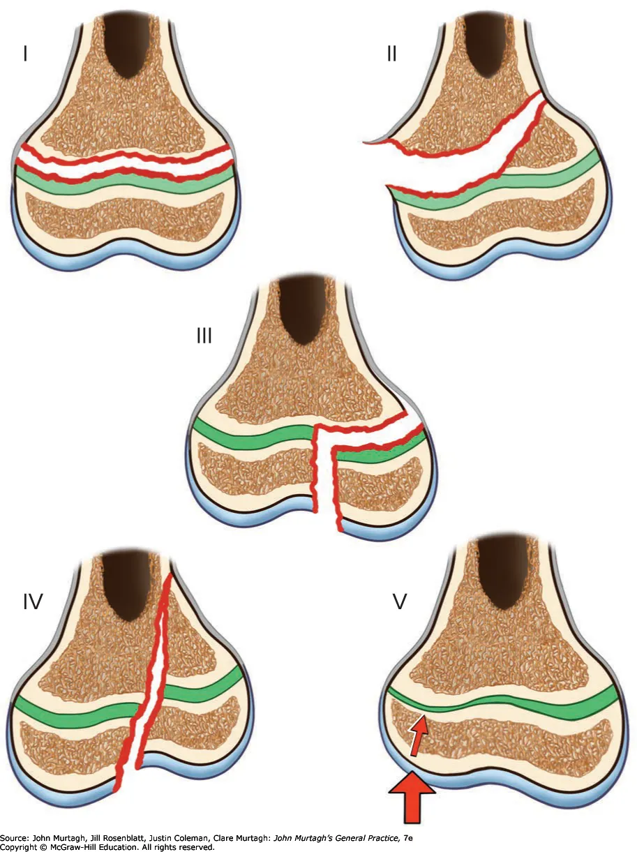
S / Straight / Slipped – I: fracture passes through the growth plate; good prognosis. A / Above – II: fracture along the growth plate then above into bone; most common (75%); good prognosis. L / Lower / Through – III: along the growth plate then down through the epiphysis; poorer prognosis; requires reduction. T / Through – IV: through growth plate, epiphysis, and metaphysis; poor prognosis; requires reduction. R / Ruined / Rammed – V: direct compression of the growth plate; uncommon (1%) but worst prognosis for growth disruption.