Knee Pain
Written 19th February 2020 - Last Update 20th August 2026
Knee pain (approach)
The regional hub for knee pain. Knee pain is common (~1 presentation per 50 patients/year); the commoner symptoms in order of frequency are pain, stiffness, swelling, clicking and locking.
Key facts
- Effusion timing is the key diagnostic clue after injury: rapid swelling (minutes to 1–4 hrs) = blood — haemarthrosis (~75% are ACL tears); swelling over 6–24 hrs to 1–2 days = synovial fluid — traumatic synovitis.
- Valgus-producing strains tend to cause ligament injuries; twisting injuries tend to cause meniscal tears.
- A ruptured ACL is a commonly missed injury — suspect it with valgus strain or sudden pivoting, a crack/pop, rapid haemarthrosis, or inability to weight-bear.
- Collateral ligament repair should be early; isolated ACL rupture can have delayed reconstruction if instability persists (early surgery mainly for high-performance athletes); combined MCL+ACL repaired early risks stiffness.
- Acute spontaneous knee inflammation may be systemic: RA, rheumatic fever, gout/pseudogout, spondyloarthropathy, Lyme disease, sarcoidosis. If infection is suspected, aspirate the joint.
- The hip and lumbosacral spine (L3–S1) refer pain to the knee — examine the joint above.
- Anterior knee pain is the commonest knee pain (≥11% of sports-related MSK problems) — usually patellofemoral pain syndrome; benign, good prognosis.
- Consider Osgood-Schlatter disorder in the 10–14-year-old (especially boys) with knee pain.
Red flags
- Acute swelling with or without trauma
- Acute (or acute-on-chronic) erythema
- Systemic features (e.g. fever) without trauma
- Unexplained chronic persistent pain
- Suspected knee dislocation — high-velocity mechanisms usually cause it, but it also occurs from low-energy mechanisms in obese patients, and up to half spontaneously reduce before assessment, so a grossly unstable post-traumatic knee (or two or more lax ligaments) should be treated as a reduced dislocation. The presence of pulses does not exclude vascular injury — check ankle-brachial index (ABI <0.9 indicates vascular insufficiency). Emergency surgery is required for vascular insufficiency, compartment syndrome, an irreducible dislocation, or an open dislocation, and a vascular injury needs repair within 8 hours for the best outcome.
Diagnostic strategy (Murtagh)
- Probability: ligamentous sprain/strain; osteoarthritis; patellofemoral syndrome; prepatellar bursitis.
- Serious, not to miss: ACL/PCL tear; DVT; septic arthritis, TB; neoplasia (primary bone tumour, metastases); rheumatoid/juvenile arthritis; rheumatic fever.
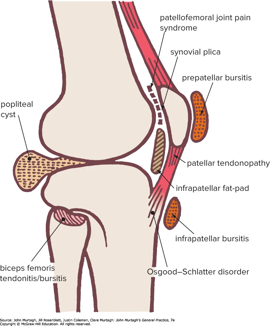
- Pitfalls: referred back/hip pain; foreign body; osteochondritis dissecans; osteonecrosis; synovial chondromatosis; Osgood-Schlatter; meniscal tears; gout; ruptured popliteal (Baker) cyst; Hoffa fat-pad syndrome.
Key questions
Injury: exact mechanism (awkward landing, direct blow and from where, twisting)? · pop/snap felt? · wobbly/unsteady, bones “separating”? · how soon pain, how soon swelling? · able to walk after, or carried off? · previous injury/surgery? · workers compensation? No injury: pain after walking/jogging? · much kneeling (carpet layers, cleaners)? · locking or catching? · swelling, grating? · rest pain/morning stiffness? · pain on stairs?
Symptom patterns
- Locking (sudden inability to fully extend, typically at 10–45°, flexion preserved) — true locking: bucket-handle meniscal tear, loose body (e.g. from osteochondritis dissecans), torn ACL remnant, articular cartilage flap, avulsed anterior tibial spine, dislocated patella, synovial osteochondromatosis. Pseudo-locking: patellofemoral disorders, grade I–II MCL tear, ACL strain, gross effusion, hamstring spasm.
- Catching — same list, but especially a subluxing patella and loose bodies.
- Recurrent swelling — patellofemoral syndrome, osteochondritis dissecans, degenerative joint disease/meniscal tears, arthritides.
- By site: anterior — fat-pad inflammation, patellofemoral syndrome, OA, patellar tendinopathy, osteonecrosis; medial/lateral — OA of that compartment, meniscal lesions, patellofemoral syndrome. Patellar tendinopathy classically causes focal tenderness at the inferior pole of the patella with pain on squatting/jumping and, like other anterior knee pain, discomfort after prolonged sitting with the knee flexed (the “movie theatre sign”).
Examination
Physical examination differentiates most causes of knee pain without imaging; MRI can usually wait for an outpatient orthopaedic review rather than being ordered urgently.
- Effusion: ballottement test (push the patella against the femur after milking fluid distally — a bounce-back is positive) and the sweep test (stroke fluid from the medial joint line up and around, then down the lateral side, watching for a wave/bulge). Oedema of the knee after trauma should raise concern for ligamentous injury.
- Ligament stability: Lachman test (ACL; sensitivity 80–99%, specificity 95–96%) is the most reliable single test — physical examination performed after the acute effusion settles is at least as accurate as MRI for diagnosing ligament tears. Anterior drawer test (ACL; less sensitive than Lachman’s), posterior drawer test/posterior sag sign (PCL), and valgus/varus stress testing at 30° flexion (MCL/LCL respectively) complete the set. Grade by laxity: grade I 1–4mm, grade II 5–9mm, grade III ≥10mm. Exam manoeuvres can be difficult in the acute setting because of pain and effusion and may need to be repeated as an outpatient once these settle.
- Meniscal tests: joint-line tenderness with the knee flexed to 90° at the edge of the table (83% sensitive, 83% specific); the Thessaly test — standing on one leg, squatting to 20° flexion while internally/externally rotating the knee (75% sensitive, 87% specific); McMurray’s test (61% sensitive, 84% specific).
- Imaging: the Ottawa knee rules help decide who needs an X-ray after acute injury with only minor findings — a systematic review of 8 studies (7,385 patients) found 99% sensitivity (49% specificity) for ruling out fracture, supporting their use to avoid unnecessary radiography.
Related pages
- Knee Fractures & Dislocations (patella, tibial plateau, tibial spine/tuberosity avulsion, and the don’t-miss knee dislocation red flag)
- Ligament injuries (ACL, PCL, MCL/LCL)
- Patellar Dislocation (reduction technique, brace vs surgery, recurrence risk)
- Acute Muscle Strains & Tears (quadriceps strain, and hamstring/calf strains presenting as posterior knee/thigh pain)
- Meniscal tears
- Patellofemoral pain syndrome
- Knee osteoarthritis
- Knee: spontaneous & idiopathic conditions (osteochondritis dissecans, osteonecrosis incl. SPONK, Osgood-Schlatter disorder)
- Tendinopathies & bursitis (patellar tendinopathy, ITB syndrome, prepatellar/anserine bursitis, Hoffa)