Patellar Dislocation
Last Update 5th September 2026
Patellar Dislocation
A routine urgent-care reduction, most often in an adolescent or young woman with a non-contact twisting injury or a direct blow to the knee. Most first-time dislocations are managed conservatively.
For collateral and cruciate ligament injuries see Ligament Injuries.
Recognition
The patella dislocates laterally in the great majority of cases. Patients describe the knee “giving way” with a pop, obvious deformity and an inability to extend the knee, followed by swelling. Adolescent girls are particularly affected, and initial episodes often follow trauma while later episodes can be atraumatic. Patellar dislocation accounts for about 2–3% of all knee injuries, an annual first-time incidence of roughly 6 per 100,000 people. The MPFL — the main static restraint of the patella — most often tears at its patellar insertion rather than in its mid-substance when the patella dislocates completely.
By the time of assessment the patella has often spontaneously relocated during extension, so a normal-looking knee on arrival does not exclude a dislocation — ask specifically whether the kneecap visibly moved out of place.
Ask about locking. Mechanical locking after a dislocation episode suggests an osteochondral loose body and needs surgical assessment rather than routine conservative management.
Test for apprehension. Passive lateral translation of the patella that produces guarding and a sense of apprehension — the apprehension sign — suggests the knee is prone to recurrent lateral dislocation. Compare with the other side.
Reduction
If the patella is still dislocated, reduce it promptly: flex the hip, apply gentle medially-directed pressure to the lateral pole of the patella, then slowly extend the knee. This can also be done with the patient sitting with the leg hanging off the edge of the bed. Analgesia or sedation first, as tolerated.
X-ray
Obtain AP, lateral and skyline (sunrise) views after any reduction — before, if the patella is still dislocated and imaging doesn’t delay reduction unduly. X-ray identifies fractures, loose bodies and malalignment; fragments most often come off the medial patellar facet or lateral femoral condyle.
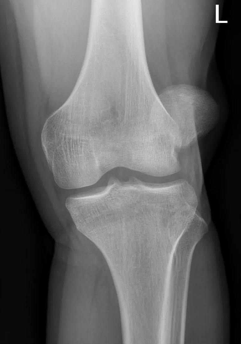
Image credit: Patellaluxation ap 002.jpg — Hellerhoff, CC BY-SA 3.0, via Wikimedia Commons (https://commons.wikimedia.org/wiki/File:Patellaluxation_ap_002.jpg).

Be wary of a large haemarthrosis with an apparently normal X-ray — this can mean a sizeable osteochondral fragment that needs early fixation and is easy to miss on plain films alone. Patellar dislocation is, in fact, the second most common cause of traumatic knee haemarthrosis.
Management
First-time dislocation without a loose body or major intra-articular injury is managed conservatively:
- Analgesia, ice and a short course of NSAIDs for pain and swelling.
- A knee brace or patella-stabilising sleeve for 2–4 weeks while soft tissue heals, allowing weight-bearing as tolerated.
- Structured physiotherapy started early, focused on quadriceps and vastus medialis obliquus strengthening, core strengthening and proprioception — this is what determines the outcome, not the brace.
There is ongoing debate about early surgical stabilisation for a first-time dislocation; the trial evidence available is not strong enough to justify routinely operating instead of the conservative pathway above.
When to refer for surgical opinion
- A first dislocation with an osteochondral fracture or loose body on imaging (locking is the clinical clue).
- MRI-confirmed disruption of the medial patellofemoral ligament (MPFL) with ongoing instability.
- Failure to improve with an adequate course of conservative management.
- Recurrent dislocation — surgical patellofemoral stabilisation is indicated when conservative care fails to control recurrent instability.
Prognosis and safety-netting
Warn every patient that re-dislocation is common — reported rates run from about 15% up to 60% after a first episode, higher again after a second — and that physiotherapy adherence over the following two to three months is what most reduces that risk. Many patients remain symptomatic for months even without re-dislocation: expect reduced sports participation and difficulty with kneeling and squatting in the first six months, and counsel accordingly before return to pivoting sport. Recurrence risk is highest in those who dislocate for the first time before age 20.