Neonatal Skin Lesions & Birthmarks
Written 8th April 2020 - Last Update 28th August 2026
Neonatal Skin Lesions & Birthmarks
See also infantile haemangioma (a fourth vascular birthmark, kept as its own page), and umbilical granuloma.
Vascular birthmarks
Salmon patch (naevus simplex)
Flat, poorly-defined patches of pink/red skin that blanch on compression — affects ~40% of newborns. Seen at the nape of the neck (“stork bite”) and between the eyes/eyelids (“angel’s kiss”). More prominent with crying, fever, breath-holding, straining, vigorous activity, or temperature change.
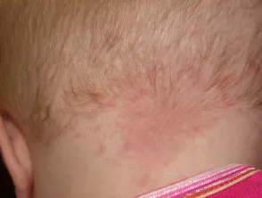
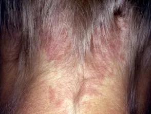
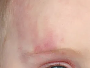
Natural history and management: fades over 6–12 months (some sources: 1–2 years) — no treatment needed. Nape-of-neck lesions fade more slowly and incompletely — up to 50% persist indefinitely into adult life. Pulsed dye laser can be considered to lighten a persistent, cosmetically bothersome lesion.
Port wine stain
A capillary vascular malformation — ~3 in 1000 neonates. Present from birth; flat at birth, can become lumpy/more unsightly over time, growing in proportion with the child. Well-defined borders — generally affects one side of the body with a sharp midline cutoff; the face is most commonly affected.
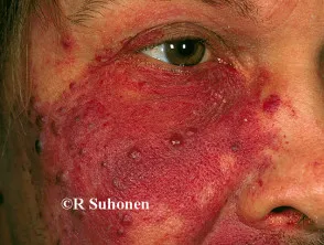
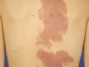
Associated vascular syndromes:
- Sturge-Weber syndrome — a port wine stain in the ophthalmic division of the trigeminal nerve is associated with glaucoma and seizures.
- Parkes Weber syndrome.
- Klippel-Trénaunay syndrome.
- Hyperkeratotic cutaneous capillary-venous malformation.
- Proteus syndrome.
Management: pulsed dye laser is the treatment of choice — cosmetic treatment should be attempted before school starts if the family wishes to pursue it. Refer for specialist opinion, and to assess for the associated syndromes above.
Lymphatic malformation (lymphangioma)
A vascular naevus/birthmark from malformed, dilated lymphatic vessels — present at birth, but can subsequently grow.
Types:
- Cystic hygroma — large fluid-filled spaces.
- Cavernous lymphangioma — can affect any body site, including the tongue; presents as a skin-coloured, red or bluish rubbery swelling under the skin.
- Lymphangioma circumscriptum — clusters of small, firm blisters resembling frogspawn.
Colour varies: skin-coloured, red/purple (if vascular involvement), or brown/black.
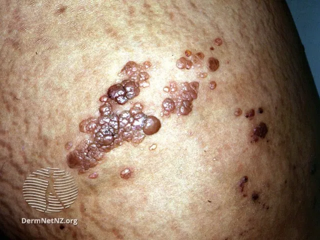
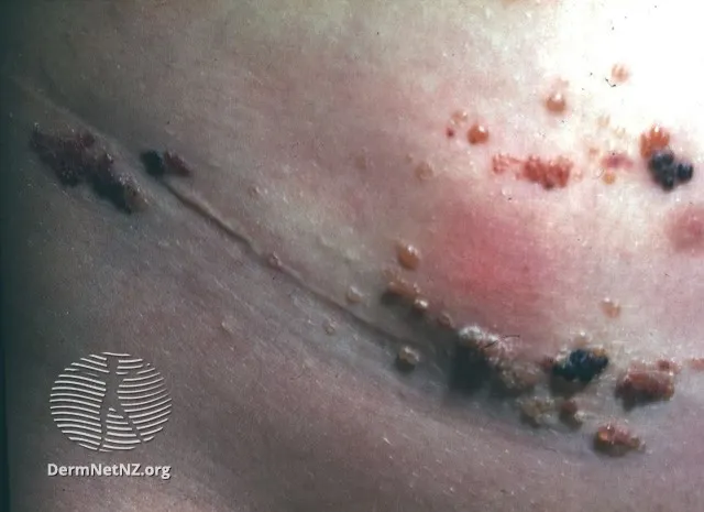
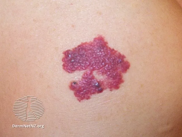
Management:
- Cystic hygroma and cavernous lymphangioma — generally removed surgically.
- Lymphangioma circumscriptum — benign, usually needs no treatment; can be removed by surgery, dermabrasion or laser if necessary.
Benign transient neonatal skin findings
Milia
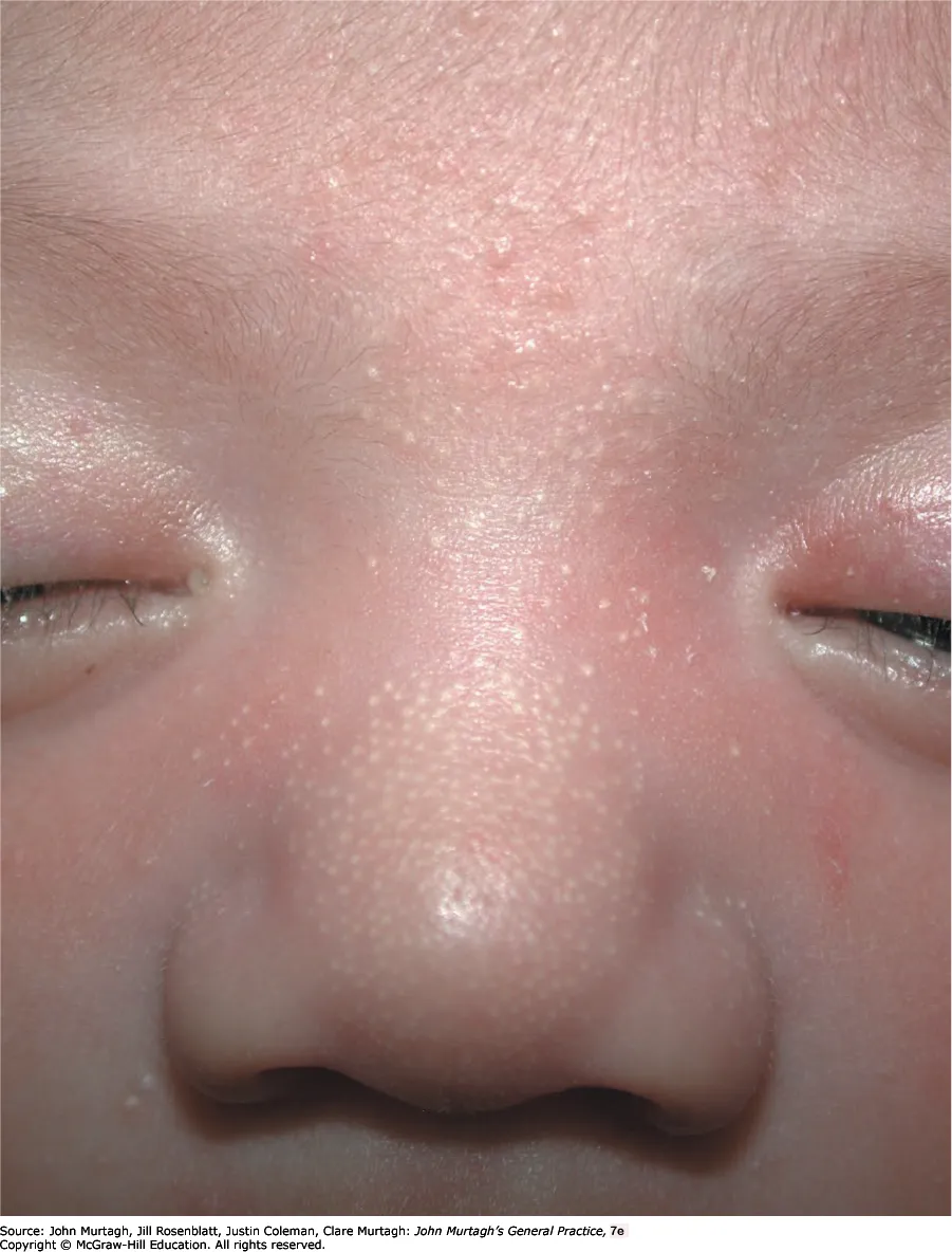
Blocked sebaceous glands producing pearly white 1–2 mm lumps under the skin, especially on the face. Disappear after several weeks — no treatment needed.
Miliaria (sweat rash)
Related to overheating — occurs in skin folds (neck, armpits) and the face. Most common in neonates (mean age ~1 week); frequent in hot, humid or tropical climates.
Types:
- Crystallina — sweat duct obstruction near the skin surface; 1–2 mm superficial clear blisters that break easily.
- Rubra (most common) — obstruction deeper in the epidermis; red, 2–4 mm, very itchy non-follicular papules/papulovesicles with background erythema.
- Profunda (tropical anhidrosis) — sweat leaks into the dermis; asymptomatic, flesh-coloured 1–3 mm papules.
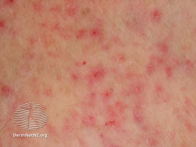
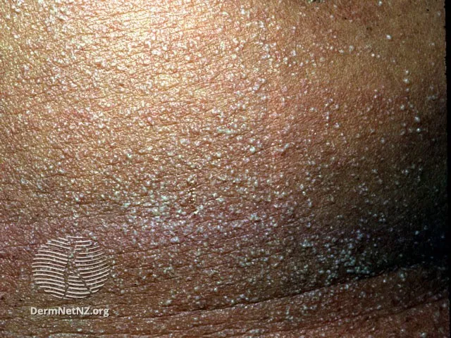
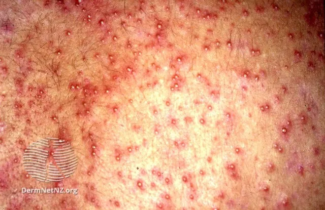
Management: benign — resolves within a day or two of a cooler environment, no treatment or complications. If problematic:
- Keep skin dry and cool (fan, air-conditioning).
- Loose-fitting cotton clothing.
- Avoid frequent bathing/overuse of soap.
- Calamine lotion, emollients.
- Mild topical steroid (e.g. hydrocortisone) if inflamed.
Toxic erythema of the newborn
A benign condition affecting ~half of babies, usually appearing 1–2 days after birth (occasionally up to 2 weeks later).
Presentation: 2–3 mm erythematous macules, papules and pustules with a surrounding blotchy erythema — a “flea-bitten” appearance. Starts on the face and spreads to the torso/limbs, sparing the palms and soles. Individual lesions rarely persist more than a day; the overall rash fades over several days. The infant is otherwise well.
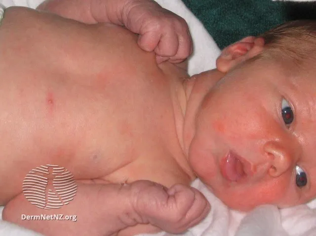
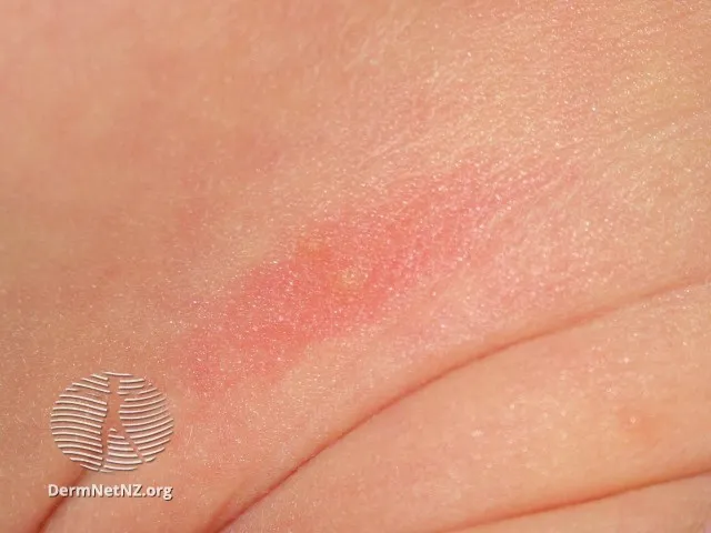
Management: no treatment required — reassurance only.
Mongolian blue spot (dermal melanocytosis)
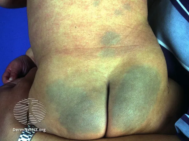
Blue-grey discolouration of the skin over the lower back and sacrum, most common in children of East Asian and other dark-skinned backgrounds. Usually disappears by age 4 — no clinical significance.
Cradle cap
Seborrhoeic dermatitis of the scalp — greasy yellow scales from sebum combining with dead skin cells.
Management:
- Mild: reassurance — resolves over weeks to months.
- More severe:
- soften with vegetable oil overnight, then gently brush off
- baby shampoo, then gentle brushing
- ketoconazole 2% (Nizoral) shampoo twice weekly.