Foot: Spontaneous & Idiopathic Conditions
Written 21st February 2020 - Last Update 25th August 2026
Foot: Spontaneous & Idiopathic Conditions
Osteochondroses of the immature midfoot/forefoot skeleton — abnormal development, injury or overuse of a growth plate/ossification centre, with no traumatic or inflammatory trigger. See also Sever disease (the same family of condition, affecting the calcaneum — kept with the Achilles-region page), osteochondritis dissecans (a related but distinct entity affecting the knee) and osteonecrosis (adult forms).
Köhler disease (navicular)
Avascular necrosis of the navicular, cause unclear; self-limiting with intermittent symptoms over 1–3 years.
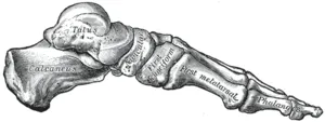
- Demographics: age 3–7 (occasionally older); boys 4×; bilateral in ~25%.
- History: midfoot pain with a limp; can be asymptomatic.
- Examination: ± swelling, warmth, redness; antalgic limp (may weight-bear on the lateral foot); point tenderness over the navicular (medial midfoot, above the 1st cuneiform, articulating with the talus).
- Investigations: X-ray shows sclerosis, fragmentation and flattening of the tarsal navicular — most reorganise after the disease runs its course (radiographs improve ~6–48 months from symptom onset); some remain deformed but almost all stay asymptomatic; no long-term disability.
- Management: NSAIDs; short-leg walking cast if pain limits activity (shortens symptom duration) — complete recovery with temporary rest, ± supportive strapping.
Freiberg disease (2nd metatarsal head)
Infarction/fracture of a lesser metatarsal head — most often the 2nd (rarely 4th/5th) — thought to follow microtrauma-related disruption of blood supply and stress overload, leading to eventual head collapse.
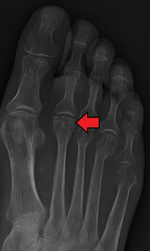
- Demographics: age 12–18 (also young adults); girls 4×; female adolescent athletes.
- History: forefoot pain, swelling and stiffness localised to the 2nd MT head; worse on weight-bearing.
- Examination: peri-articular swelling and tenderness; limited 2nd MTP motion.
- Investigations: X-ray — subchondral sclerosis early, flattening of the MT head, joint destruction late.
- Management:
- early disease: activity limitation, NSAIDs, short-leg walking cast/boot 4–6 weeks, stiff-soled shoe with metatarsal bar/pad
- refer for surgery if conservative management fails.