Achilles Disorders
Written 22nd February 2020 - Last Update 25th August 2026
Achilles Disorders
Tendinopathy, rupture and bursitis of the largest, strongest tendon in the body. (Consolidates the former Achilles Tendinopath” and “Achilles Bursitis” notes.) “Injuries are bimodal: acute ruptures in the younger/active, chronic overload conditions with age.
Achilles tendinopathy
Degenerative ± inflammatory change in the tendon or surrounding structures; overuse, dorsiflexor/plantarflexor imbalance, poor tendon blood supply, genetics, fluoroquinolone antibiotics, and inflammatory arthropathy all contribute.
- Non-insertional (mid-tendon, 2–6 cm above the calcaneus — the watershed zone of poor vascularity): younger, fitter, active patients (annual incidence 7–9% in high-level runners). Gradual-onset mid-tendon pain with fusiform swelling.
- Insertional: middle-aged, overweight/unfit, diabetes — often a combination of retrocalcaneal bursitis, Haglund deformity and calcific deposits at the insertion (see bursitis below).
- History: tendon pain during and after weight-bearing activity, worse in the morning.
- Examination: tenderness ± thickening/swelling of the tendon; pain on single-leg heel raise or hopping. Always check for rupture (Thompson test).
- Investigations: clinical diagnosis; ultrasound if unclear; X-ray if chronic (Haglund deformity, calcification).
- Management:
- reduce load (e.g. running volume) then graded reintroduction; weight loss; heel raises/footwear modification
- physiotherapy/podiatry-guided loading program — isometric then progressive strengthening (eccentric calf loading) is the evidence-based cornerstone; non-operative management succeeds in 65–90%
- do NOT inject corticosteroid — risk of tendon rupture; PRP/GTN patches/prolotherapy have a limited evidence base
- sport & exercise physician or orthopaedic referral if failing 6 months of conservative care (FHL tendon transfer is reserved for severe cases).
Achilles tendon rupture
- Demographics: bimodal — 25–40-year-old recreational athletes (acceleration–deceleration mechanism, up to 90%) and >60s with low-energy mechanisms on a degenerative tendon. Men >2×. Risk factors: corticosteroids, fluoroquinolones (latency 2–60 days), RA, gout, ankylosing spondylitis.
- History: sudden “gunshot”/hit-in-the-back-of-the-heel sensation, falls over, bruising/swelling, difficulty walking (especially on toes). Up to 25% are missed at first presentation — patients can often still weakly plantar flex and pain may be mild.
- Examination — Simmonds’ triad (prone or kneeling on a chair):
- Look — increased angle of declination (foot rests in more dorsiflexion than the other side)
- Feel — palpable gap
- Move — calf squeeze test (Simmonds/Thompson): no plantar flexion = positive. Compare sides. Any two of three ≈ 100% sensitive.
- Beware the calcaneal tuberosity avulsion fracture (elderly, diabetes, immunosuppression, osteopaenia): a bony lump on palpation or tented skin — plantarflexed cast + urgent X-ray; this is a surgical emergency (skin necrosis risk).
- Investigations: clinical diagnosis; ultrasound (better than MRI for gap/partial-vs-complete) if in doubt; X-ray to exclude avulsion.
- Management:
- immobilise in plantarflexion (backslab or CAM boot with heel wedge) as early as possible — ideally within hours — to prevent the tendon healing long (functional lengthening → weakness)
- crutches, light weight-bearing; orthopaedic/fracture-clinic review within 7 days
- supervised non-operative management with an accelerated functional rehabilitation protocol now achieves outcomes equivalent to surgery (RCT: no difference in strength/ROM at 2 years; surgery halves re-rupture ~12%→5% but has ~30% vs 8% complication rate). Boot > cast; early weight-bearing improves outcomes. Typical protocol: boot with 3 cm heel raise, wean wedges from week 6, out of boot from week 12, then graded strengthening/return to sport
- DVT/PE risk is elevated (~2.7%/~1.8%) — no routine chemoprophylaxis, but educate about calf pain/dyspnoea and encourage weight-bearing.
Achilles tendon bursitis
Two bursae: posterior/superficial (skin–tendon; classically young women, shoe friction) and deep/retrocalcaneal (calcaneus–tendon; associated with Haglund deformity, a posterosuperior calcaneal prominence).
- Examination: superficial — visible tender swelling with erythema; deep — squeeze in front of the tendon with thumb and index finger: tender bulging either side of the tendon; pain on dorsiflexion.
- Investigations: clinical; lateral X-ray shows Haglund deformity; ultrasound differentiates bursitis from tendinosis.
- Management: avoid shoe heel pressure (podiatry can modify work boots), 1–2 cm in-shoe heel raise, NSAIDs, calf/tendon stretching then strengthening once settled. Avoid corticosteroid injection (rupture risk). Refractory: surgical retrocalcaneal bursa excision + Haglund resection ± tendon debridement.
Sever disease (calcaneal apophysitis, paediatric)
A different mechanism from the adult conditions above — a traction apophysitis of the calcaneal growth plate at the Achilles insertion, seen in growing children rather than adults.
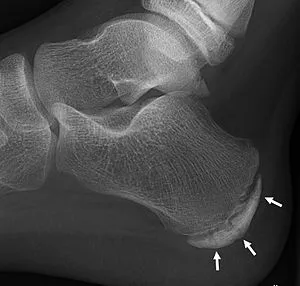
- Demographics: boys, age 7–15 (average 10); running/jumping sports (immature athletes).
- History: painful, tender heel at the Achilles insertion; tight Achilles tendon.
- Examination: positive squeeze test (pain with medial-lateral compression over the calcaneal tuberosity); tenderness over the calcaneal apophysis.
- Investigations: X-ray mainly to exclude other causes (osteomyelitis, bone cysts) — may show apophyseal sclerosis/fragmentation, but has low sensitivity/specificity (diagnosis is largely clinical).
- Management:
- avoid flat-heeled shoes (a slight heel is beneficial); heel cups/pads
- activity modification — restrict strenuous sport for ~12 weeks, then review
- Achilles stretches (may reduce recurrence), ice before/after sport, NSAIDs
- short leg cast for persistent pain.
- Self-limiting — resolves with apophyseal maturation/closure.