Syncope
Written 24th February 2020 - Last Update 27th August 2026
Syncope
Murtagh groups fits, faints and funny turns” together — a small amount of history taking usually suffices to differentiate between them, though this isn’t always possible in the elderly with unwitnessed events. “In syncope there is usually a prodrome (light-headedness, nausea, sweating, loss of vision/hearing) followed by collapse and rapid return of consciousness — the prodrome may be terminated by lying down. A brief or absent prodrome suggests cardiac syncope, which usually needs hospital admission for monitoring (though this can sometimes be done as an outpatient if the patient is stable with a normal ECG).
Syncope accounts for 1–3.5% of emergency department presentations and around 6% of hospital admissions in the US, and most episodes are benign — vasovagal (neurocardiogenic) mechanisms alone account for close to half of all syncope. Cardiac causes become proportionally more common with age, while non-cardiac (especially vasovagal) mechanisms predominate in young adults.
Causes of syncope
- Cardiac syncope (< 5% of cases).
- Vasovagal syncope (common) — needs a trigger (emotional distress, posture, situational triggers such as micturition or neck movement); associated with hypotension and bradycardia from vagal outflow. First-line management is conservative — avoiding known triggers, tilt training, and increasing salt and fluid intake; if this fails, drug options that may be considered include beta-blockers, SSRIs, fludrocortisone or midodrine.
- Orthostatic hypotension — occurs within 3 minutes of standing, with a documented systolic BP drop; more common in older adults from dehydration, drugs (e.g. antihypertensives), or autonomic failure (Parkinson’s disease, diabetes). Management starts with rising slowly from supine/sitting, reviewing and where possible stopping culprit drugs (diuretics, vasodilators), and compression stockings/IV fluids for volume depletion. Midodrine — an alpha-1 agonist prodrug causing venous vasoconstriction — may be considered for refractory, severe symptomatic cases once other treatable causes are addressed; start at a low dose and increase weekly, taking the evening dose ≥4 hours before bedtime to limit supine hypertension (roughly 1 in 8 treated patients develops it). Evidence for midodrine is modest: trials show it raises standing systolic BP, but a systematic review found no clear benefit on the supine-to-standing BP drop itself, and its Australian approval is limited to severe symptomatic autonomic-dysfunction cases after exacerbating factors are addressed and other treatments have failed. Contraindicated if creatinine clearance <30 mL/min.
- Infections causing vasodilation and dehydration.
- POTS (postural orthostatic tachycardia syndrome) — a multi-system disorder of
orthostatic intolerance with a marked rise in heart rate on standing but without
the orthostatic BP drop that defines orthostatic hypotension, so it does not itself
cause true syncope — it instead causes pre-syncope-like symptoms (recurrent
light-headedness, palpitations, fatigue without loss of consciousness). Most
patients are young women (up to 80% female; typical onset age 14, mean 21±12
years), and it commonly co-exists with joint hypermobility, fibromyalgia and
migraine.
- Diagnostic criteria (2015 Heart Rhythm Society consensus, all four required): sustained HR rise ≥30bpm (≥40bpm if aged 12–19) within 10 minutes of standing; no orthostatic BP drop ≥20/10mmHg; orthostatic-intolerance symptoms worse upright with rapid improvement lying down, present >6 months; no other cause of sinus tachycardia.
- Three overlapping pathophysiological subtypes: neuropathic (impaired sympathetic venoconstriction, mainly lower-limb), hypovolaemic (low plasma volume, urine sodium <100mmol/24h), and hyperadrenergic (standing systolic BP rise >10mmHg, plasma noradrenaline >600pg/mL).
- Initial work-up: orthostatic vital signs, 12-lead ECG, 24-hour Holter (to exclude arrhythmia), and bloods (FBC, iron studies, active B12, electrolytes, renal function, TFTs, plasma noradrenaline, morning cortisol) to screen other causes of tachycardia; a tilt table test can confirm postural HR variability.
- Management is non-pharmacological first (graded exercise reconditioning, sodium intake >10g/day with ≥2.5L fluid/day, class 2 compression garments above 30mmHg), then pharmacological by subtype: beta-blockers (e.g. propranolol 10–40mg tds, bisoprolol 2.5–5mg bd, metoprolol 25–100mg daily, atenolol 12.5–50mg daily) or ivabradine 2.5–5mg bd to reduce sinus tachycardia; midodrine 2.5–10mg bd or clonidine 0.2–0.6mg bd to enhance vaso/venoconstriction (clonidine favoured for the hyperadrenergic phenotype); pyridostigmine 30–60mg bd/tds for a suspected neuropathic phenotype; fludrocortisone 0.1–0.2mg daily or desmopressin 0.1–0.4mg bd to expand blood volume (hypovolaemic phenotype).
- Pulmonary embolism.
Evaluation approach
Featuring the 2017 US Syncope Guidelines, HealthPathways, Primary Care RAP, and LITFL.
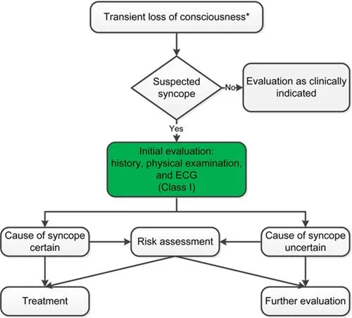
The cornerstone of syncope evaluation is history, examination, and ECG — this risk-stratifies the patient into needing hospital admission for cardiac monitoring, outpatient evaluation, or no further evaluation at all.
Assessment
History — first-hand witness accounts are invaluable. Ask about: circumstances (sitting/standing/lying); warning symptoms; tongue biting (suggests seizure); confusion afterward; speed of recovery; hyperventilation beforehand (suggests anxiety); chest pain; shortness of breath; palpitations.
Cardiac syncope red flags: exertional syncope; syncope while supine; known heart disease or conduction problems; first syncope in an older person without postural hypotension; family history of sudden death (<40yo); abnormal ECG; persistently abnormal vital signs.
Examination: lying and standing BP; cardiovascular exam for murmurs; basic neurological exam.
Investigations
ECG is mandatory in all patients (see ECG Interpretation for a systematic reading approach). The rest of the work-up is guided by the history/examination findings above.
Consider CT brain if: suspected first seizure; secondary trauma (hit head on the ground); suspected TIA/stroke; a neurological deficit; age > 65; sudden-onset headache; on warfarin.
Choosing Wisely (IMSANZ): don’t request Holter monitoring, carotid duplex, echocardiography, EEG, or telemetry for a first presentation of uncomplicated syncope with no high-risk features. These tests have very low diagnostic yield unless there are features/risk factors for: arrhythmia (palpitations preceding syncope, exertional or unheralded syncope, a history suggestive of heart failure/ischaemic heart disease); carotid stenosis (syncope with focal neurological symptoms suggestive of TIA); cardiac valvular disease (a definite murmur); or seizures (very rarely presents as syncope without other epileptic features — tongue biting, urinary incontinence, post-ictal confusion, muscle pain).
Most syncope is vasovagal or from postural hypotension, for which careful history plus lying/standing BP, combined with a standard 12-lead ECG, are the most important diagnostic tools.
Tilt table testing is useful when syncope is recurrent with an unclear cause and no cardiac disease, when vasovagal syncope is suspected but life-threatening causes need excluding first, or to distinguish reflex syncope from orthostatic-hypotension syncope.
Risk stratification, disposition and management
Disposition is often the hardest part of managing syncope — admission rates vary widely between health systems (around 80% of ED presentations in the US, versus as low as 10% in Canada), a gap that likely reflects differing thresholds for chasing rare but serious causes rather than a real difference in patient acuity. Numerous risk-scoring pathways (e.g. the Boston Syncope Criteria, based on known cardiac disease, associated symptoms like chest pain/dyspnoea/palpitations, and ECG abnormalities) have been published to standardise this, but none clearly outperforms a thorough history and examination — a patient with no relevant comorbidities and an unremarkable ED workup is generally safe for discharge.
For recurrent syncope of uncertain origin without high-risk features, an implantable loop recorder (ILR) — a small subcutaneous device that continuously records and loops the ECG — is recommended early in the diagnostic pathway (with high-risk patients getting one after an initial negative work-up). A Cochrane review of 4 RCTs (579 adults) found moderate-quality evidence that an ILR-based strategy increases the rate of an aetiological diagnosis compared with conventional work-up, with a trend toward fewer syncope recurrences once a diagnosis is reached and a lower cost per diagnosis despite higher up-front costs; there was no evidence of a mortality difference (low-quality evidence) and quality-of-life data were inconclusive.
General acute management: get the patient supine with legs elevated as soon as possible to aid recovery from reflex hypotension, keep them lying down and prevent early standing, and assess for injury from the fall itself. Safety-net advice: patients should not drive or operate heavy machinery until their work-up (or GP follow-up) is complete. Prognosis is generally good for syncope of unknown cause without underlying heart disease; those with known cardiac disease need regular GP and cardiology follow-up, since annual mortality is markedly higher with a cardiac cause (reported 18–33%) than a non-cardiac one (0–12%). Consider an underlying mental health or substance-use contribution when the cause remains unclear.
ECG pattern recognition in syncope
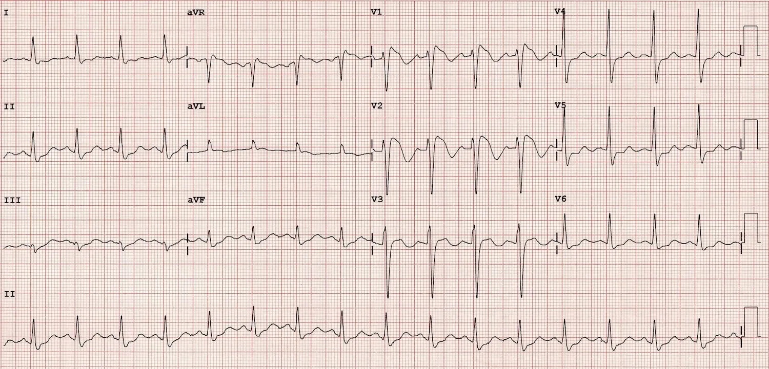
Mnemonic — “Can Quick BRAD Walk Home”: Conduction blocks, QT syndrome (long/short), Brugada syndrome, Right ventricular infarction, Arrhythmogenic right ventricular cardiomyopathy, Dilated cardiomyopathy, WPW, HOCM/LVH from aortic stenosis.
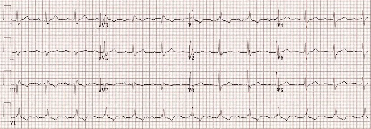
(High-grade) conduction blocks
Complete heart block, or trifascicular block (an indication for pacemaker if symptomatic) — usually RBBB with left axis deviation (from left anterior fascicular block).
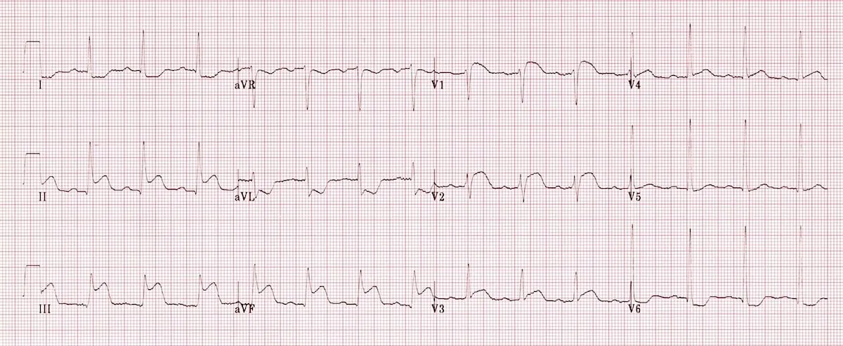
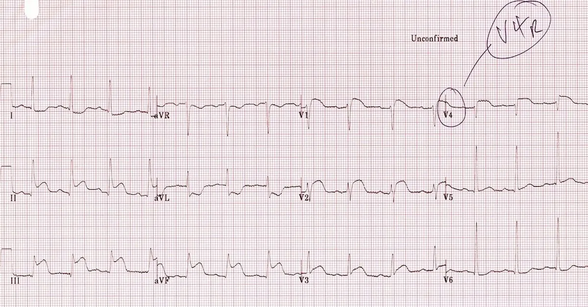
Right ventricular infarction
Uncommon in isolation; common alongside inferior infarcts. ST elevation in V1 with ST depression in V2 — consider in any inferior STEMI, and obtain right-sided leads.
Long QT syndrome
Congenital or drug-induced. Consider if QTc > 450ms (men) or > 460ms (women) — a QT nomogram is also available via Therapeutic Guidelines. Measure QT across 6 leads, take the median, and plot against heart rate.
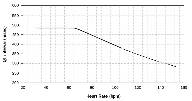
If the point falls above the nomogram’s risk line, there’s a risk of torsades — see Prolonged QT / Torsades de Pointes for management.
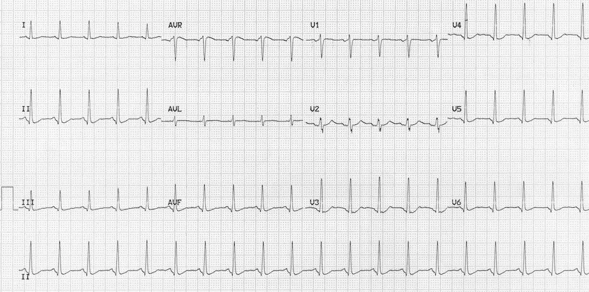
Short QT syndrome
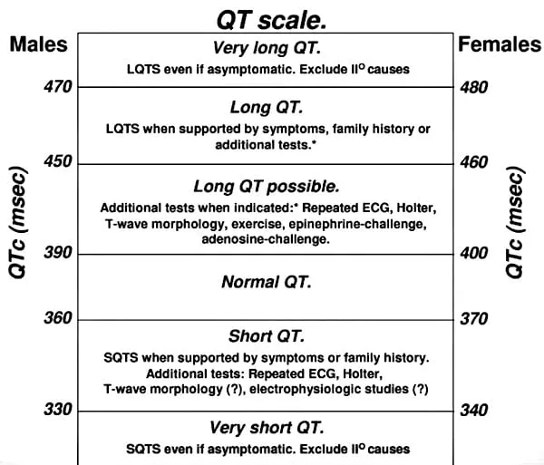
Consider if QTc < 360ms (men) or < 370ms (women) — a lack of normal QT-duration change with heart rate. Associated with rapid ventricular arrhythmias.
Brugada syndrome
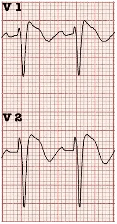
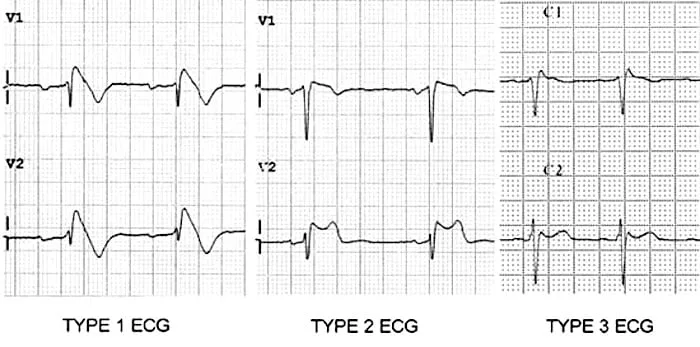
Coved ST elevation followed by a negative T-wave in V1–V2 — easy to recognise in its Type 1 form.
Arrhythmogenic right ventricular cardiomyopathy (ARVC)
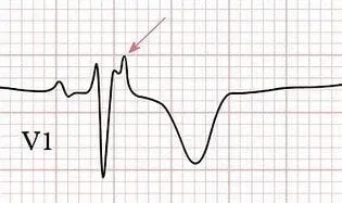
The 2nd most common cause of sudden cardiac death in young people (after HOCM); an inherited disorder. Look for an epsilon wave in V1 (a positive deflection buried at the end of the QRS).
Wolff-Parkinson-White (WPW) syndrome
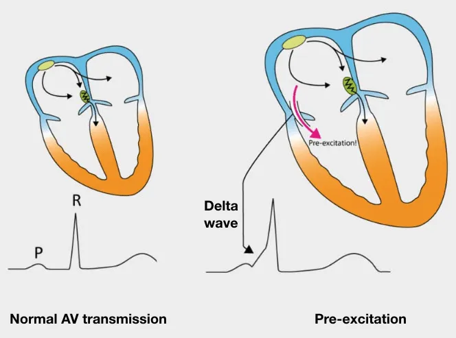
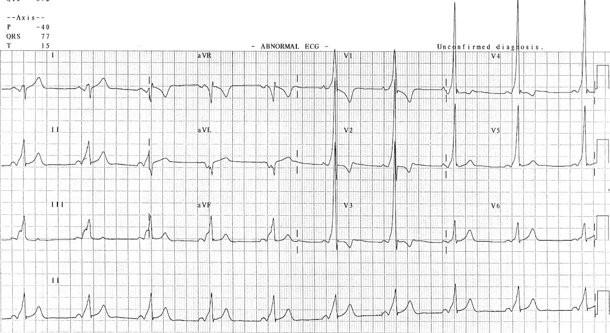
A pre-excitation syndrome causing tachyarrhythmias via conduction through an accessory pathway — see SVT & WPW for the full clinical picture. In sinus rhythm: a short PR interval and a delta wave (slurred QRS upstroke).
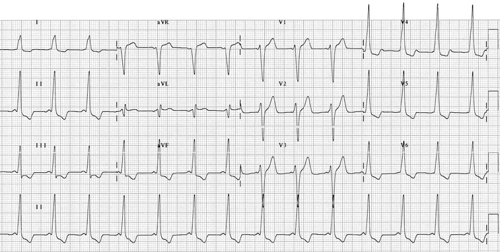
Hypertrophic obstructive cardiomyopathy (HOCM)
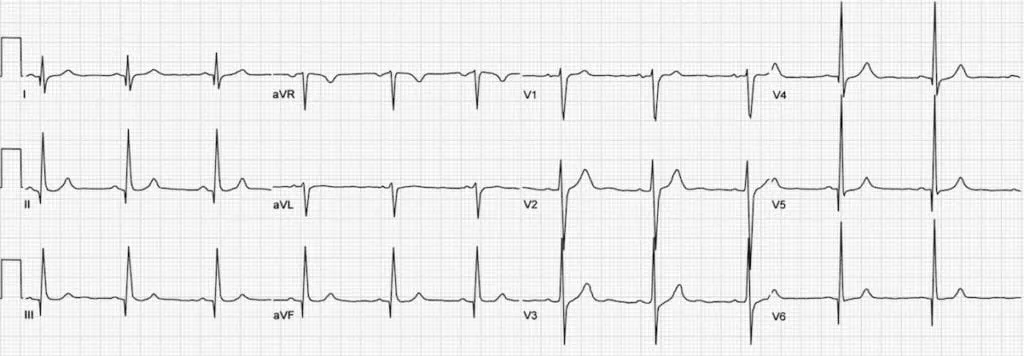
The most common inherited cardiac disorder (1:500) and the leading cause of sudden cardiac death in athletes; annual mortality 1–2%. Causes exertional syncope via arrhythmia or LVOT obstruction — see Cardiomyopathies for full management detail. ECG: dagger-like Q-waves in the lateral and inferior leads (V5/V6, I/aVL, II/III/aVF).
Quick ECG summary
- Rate/rhythm: tachy- or bradyarrhythmias.
- Intervals: short PR with delta wave (WPW); short or long QTc.
- QRS: dagger Q-waves (HOCM); LVH by voltage criteria.
- ST segment/T-waves: ischaemia; epsilon wave (ARVC); coved ST in V1–V2 (Brugada).