Cardiomyopathies
Written 27th February 2020 - Last Update 6th September 2026
Cardiomyopathies
- Dilated cardiomyopathy — dilated chamber with impaired systolic function.
- Hypertrophic cardiomyopathy — thickened ventricular wall with impaired relaxation.
- Restrictive cardiomyopathy — abnormally stiffened myocardium (fibrosis or infiltration) with impaired relaxation.
Dilated cardiomyopathy
Causes:
- Idiopathic; familial.
- Inflammatory — myocarditis, connective tissue disease, peripartum cardiomyopathy, sarcoidosis.
- Toxic — alcohol, chemotherapy (e.g. doxorubicin), methamphetamine.
- Metabolic — hypothyroidism, hypocalcaemia/hypophosphataemia.
- Neuromuscular — muscular dystrophy.
Management: as for HFrEF — see Heart Failure with Reduced Ejection Fraction.
Methamphetamine-associated cardiovascular conditions (MACCs)
Episodic and chronic methamphetamine use is linked to dilated-phenotype cardiomyopathy, takotsubo cardiomyopathy, pulmonary hypertension, accelerated coronary artery disease, myocardial infarction, systemic hypertension, hypertensive emergency, arterial vasospasm, aortic dissection, stroke, ventricular arrhythmia and sudden cardiac death. These conditions are the leading cause of natural death among people who use methamphetamine, with deaths increasing seven-fold in Australia between 2009 and 2020.
Screening
Hold a low threshold for resting ECG screening in anyone with past or present methamphetamine use. Some MACCs can cause death years after achieving abstinence.
Investigating suspected MACCs
Work-up for any suspected MACC follows the corresponding standard clinical guideline. There is a low threshold for transthoracic echocardiography in symptomatic patients.
Management
Abstinence is vital to reverse, arrest or slow progression of MACCs. Methamphetamine-associated cardiomyopathy is managed with guideline-directed medical therapy for heart failure, with cardiology input.
Harm reduction
Harm-reduction counselling is important for patients not ready to change their pattern of use:
- Advise regular breaks in use, since repeated dosing over days amplifies cardiovascular effects.
- Inform patients that smaller amounts, used less often, may cause less cardiovascular harm.
- Address modifiable cardiovascular risk factors: smoking, hypertension, hypercholesterolaemia, chronic kidney disease.
- Teach the symptoms and signs of stroke and acute coronary syndrome, and the overdose risk of co-using methamphetamine with CNS and respiratory depressants (eg opiates, sedative medication, alcohol).
- Any amount of methamphetamine use carries risk, and even small doses have been associated with death.
Hypertrophic cardiomyopathy (HCM)
An inherited disorder, with population-based studies suggesting a prevalence of up to 1 in 200 — making it the commonest known inherited cardiovascular condition, and more common than the traditionally-quoted 1 in 500 figure. Hypertrophy is not caused by chronic pressure overload. Causes exertional syncope and sudden cardiac death (SCD) — the leading cause of SCD in young athletes — via dynamic outflow obstruction or life-threatening ventricular arrhythmia. Can develop at any age; patients with a family history should have 2-yearly ECG surveillance for emerging hypertrophy.
Demographics: typical age at presentation is the mid-20s; annual mortality 1–2%.
History: family history of sudden cardiac death or unexplained drowning; exertional syncope; exertional dyspnoea.
Examination: S4; a crescendo-decrescendo systolic murmur that gets louder with Valsalva (the opposite of most systolic murmurs, reflecting dynamic outflow obstruction).
Investigations: ECG — classically dagger-like Q waves in the lateral and inferior leads (V5/V6, I/aVL, II/III/aVF).
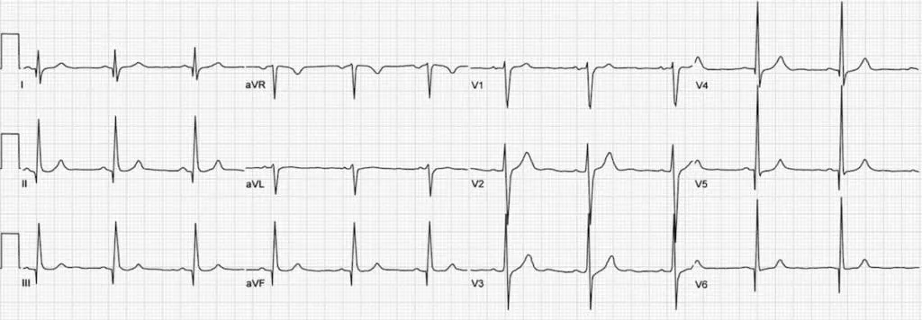
Echo shows hypertrophy and (in obstructive HCM) outflow tract obstruction.
Management of left ventricular outflow tract obstruction (LVOTO)
Stepwise per the 2023 ESC cardiomyopathy guideline:
- Non-vasodilating beta-blockers, titrated to maximum tolerated dose — first-line.
- If ineffective/contraindicated: verapamil (40mg three times daily, up to 480mg/day — use cautiously in severe obstruction, can provoke pulmonary oedema) or diltiazem (60mg three times daily, up to 360mg/day).
- Disopyramide, added to a beta-blocker (or verapamil/diltiazem if a beta-blocker isn’t possible), titrated to a maximum of 400–600mg/day — monitor QTc (reduce dose if >500ms); avoid in glaucoma, prostatism, or with other QT-prolonging drugs.
- Mavacamten (a cardiac myosin ATPase inhibitor) — TGA-approved and PBS-subsidised (2025) as second-line therapy, added to a beta-blocker (or verapamil/diltiazem) when beta-blockers/calcium antagonists/disopyramide are ineffective or poorly tolerated, before septal reduction therapy. Reduces LVOT gradient and improves symptoms/exercise capacity (EXPLORER-HCM trial); ~70% of patients show significant clinical/echo improvement at 6 months in early Australian real-world data. Requires echocardiographic monitoring of LVEF during up-titration; don’t combine with disopyramide.
- Septal reduction therapy (surgical myectomy or alcohol septal ablation) if still symptomatic.
Sudden cardiac death risk stratification
ESC 2023 recommends formally estimating 5-year SCD risk (the validated HCM Risk-SCORE calculator, incorporating age, maximal wall thickness, left atrial size, LVOT gradient, family history of SCD, non-sustained VT, and unexplained syncope) at first evaluation and re-evaluating every 1–2 years or with any change in clinical status — a more structured approach than a simple checklist of risk factors.
- ≥6% 5-year risk — ICD implantation recommended, after detailed clinical assessment.
- 4–<6% 5-year risk — ICD considered, individualised to the patient.
- Additional factors that can tip a borderline decision toward ICD: extensive late gadolinium enhancement (≥15%) on cardiac MRI, or LVEF <50%.
General measures: avoid strenuous exercise and competitive sport.
Genetics
HCM and dilated cardiomyopathy are the archetypal monogenic (“Mendelian”) cardiac conditions — usually autosomal dominant, so a first-degree relative of an affected person has a 50% chance of also carrying the variant. Rarity of a variant, its location in a functionally critical gene region, and co-segregation with disease in the family all help determine pathogenicity, but this classification is best done by a clinical geneticist — genetic testing mainly adds value for cascade testing of at-risk relatives once a proband is confirmed, similar to the cascade-screening approach already used for familial hypercholesterolaemia.
Restrictive cardiomyopathy
Abnormally rigid, but not thickened, myocardium.
Causes:
- Non-infiltrative — idiopathic, scleroderma.
- Infiltrative — amyloidosis (the most common cause overall), sarcoidosis.
- Storage disease — haemochromatosis, glycogen storage disease.
- Endomyocardial disease — radiation therapy, endomyocardial fibrosis, hypereosinophilic syndrome, metastatic tumour.
Presentation: signs of heart failure (often HFpEF).
Management: treat the underlying condition — e.g. phlebotomy for haemochromatosis, chemotherapy for AL (primary) amyloidosis.
Cardiac amyloidosis — diagnostic pathway
Amyloidosis is the most important restrictive-cardiomyopathy cause to actively consider, since it’s now both detectable non-invasively and treatable — around 95% of cases are ATTR (transthyretin, wild-type or hereditary) or AL (light chain) amyloid, and distinguishing between them changes both prognosis and treatment.
Consider amyloidosis in patients with unexplained LV wall thickening (including a historical HCM label), HFpEF (especially in older men), or low-flow low-gradient aortic stenosis (present in up to 16% of AS patients referred for TAVI). Extra- cardiac “red flags” suggesting systemic ATTR disease: bilateral carpal tunnel syndrome (up to 50% of ATTR patients), biceps tendon rupture, spinal canal stenosis, autonomic neuropathy (orthostatic hypotension, sexual dysfunction, sweating abnormality), and alternating diarrhoea/constipation. Cardiac red flags: down- titration of antihypertensives in a previously hypertensive patient, low QRS voltage on ECG discordant with echo wall thickness, persistently elevated troponin, and a disproportionately elevated NT-proBNP.
Non-invasive diagnosis is now possible, avoiding biopsy in most cases:
- Bone scintigraphy (technetium-labelled bisphosphonate tracer, e.g. PYP/HDP) — myocardial uptake grade ≥2 (visual, compared to rib uptake), combined with exclusion of a monoclonal protein (serum/urine protein electrophoresis + immunofixation, plus serum free light chains), gives close to 100% specificity and positive predictive value for ATTR-CA.
- If the monoclonal-protein screen is positive (or scintigraphy is negative), further work-up for AL amyloidosis and/or referral to a specialist amyloidosis centre for tissue diagnosis (endomyocardial, or an “off-target” biopsy — bone marrow, fat pad, or salivary gland) is needed.
- All patients with confirmed ATTR-CA should have genetic testing to distinguish wild-type from hereditary (variant) disease.
- Cardiac MRI is a useful complementary tool for structural/functional assessment and tissue characterisation, though it doesn’t replace scintigraphy/biomarker screening for subtyping.
Treatment is now disease-modifying and targeted for ATTR-CA — tafamidis (a transthyretin stabiliser) is becoming available on the PBS in Australia; diflunisal is accessible via the Special Access Scheme through Australian Amyloidosis Network centres. Refer confirmed or suspected cases for multidisciplinary, specialist-centre care.