Supraventricular Tachycardia & Wolff-Parkinson-White Syndrome
Written 24th February 2020 - Last Update 21st August 2026
Supraventricular Tachycardia & Wolff-Parkinson-White Syndrome
Supraventricular tachycardia (SVT)
Can be paroxysmal or sustained; a narrow QRS complex (<120ms) with a rate typically 150–220/min. At least 8 different types exist, with differing risk profiles and treatment responses — the two most common mechanisms are atrioventricular nodal re-entrant tachycardia (AVNRT, a reentry circuit contained within the AV node) and atrioventricular reciprocating tachycardia (AVRT, using an accessory pathway, as in WPW), which together account for the large majority of paroxysmal SVT (PSVT). AVNRT alone has an incidence of around 35 per 10,000 person-years and is the most common tachyarrhythmia in young adults; women have roughly double the risk of men, and older individuals a roughly five-fold higher risk than younger ones.
Paroxysmal SVT commonly presents with sudden onset in otherwise healthy young people — passing copious dilute urine after an attack is characteristic, thought to result from atrial natriuretic factor released by the atria in response to atrial stretch during the tachycardia. Look for predisposing factors such as an accessory pathway or thyrotoxicosis; common lifestyle triggers to ask about (and to advise limiting) include caffeine, alcohol and stimulant use, plus dehydration. Approximately 60% are due to AV node re-entry and 35% to accessory pathway tachycardia (e.g. WPW). Look for evidence of an accessory pathway after reversion, since these carry a risk of sudden death (avoid AV-nodal blocking drugs — digoxin, verapamil, diltiazem, beta-blockers and adenosine — if there is any possibility of atrial fibrillation or flutter in a WPW patient, since blocking the AV node can leave the accessory pathway to conduct the atrial rate straight to the ventricles, precipitating ventricular fibrillation — see the WPW section below). Consider sick sinus syndrome in a patient with SVT and dizziness.
Wolff-Parkinson-White (WPW) syndrome
The structural basis of SVT in WPW is an accessory pathway bypassing the AV node. A typical ECG shows a short PR interval and a slurred QRS upstroke (delta wave). WPW is uncommon — population studies put the prevalence of the ECG pattern at around 1–3 per 1,000 people — and most patients with the pattern never develop an arrhythmia at all: roughly 65% of adolescents and 40% of adults over 30 with the ECG pattern are asymptomatic, with the pattern typically discovered incidentally. Patients are prone to sudden PSVT attacks; up to 30% develop atrial fibrillation or flutter, which is the setting that actually carries the risk of sudden death in WPW (population studies estimate sudden cardiac death at roughly 0.0002–0.0015 per patient-year overall) — risk is higher with male sex, age under 35, a prior history of atrial fibrillation or AVRT, multiple accessory pathways, a septal pathway location, or a pathway capable of rapid antegrade conduction. Even one episode of PSVT warrants consideration for radiofrequency ablation — the 2015 ACC/AHA/HRS guideline recommends catheter ablation as first-line therapy for any symptomatic patient, given its high success rate and low complication rate.
For an asymptomatic patient with an incidental WPW pattern on ECG, referral for cardiology or electrophysiology review is still reasonable to discuss risk stratification (exercise testing, ambulatory ECG monitoring, or an electrophysiology study) rather than immediate treatment — an abrupt, clear loss of pre-excitation at faster heart rates suggests a low-risk pathway that can usually be managed with watchful monitoring alone.
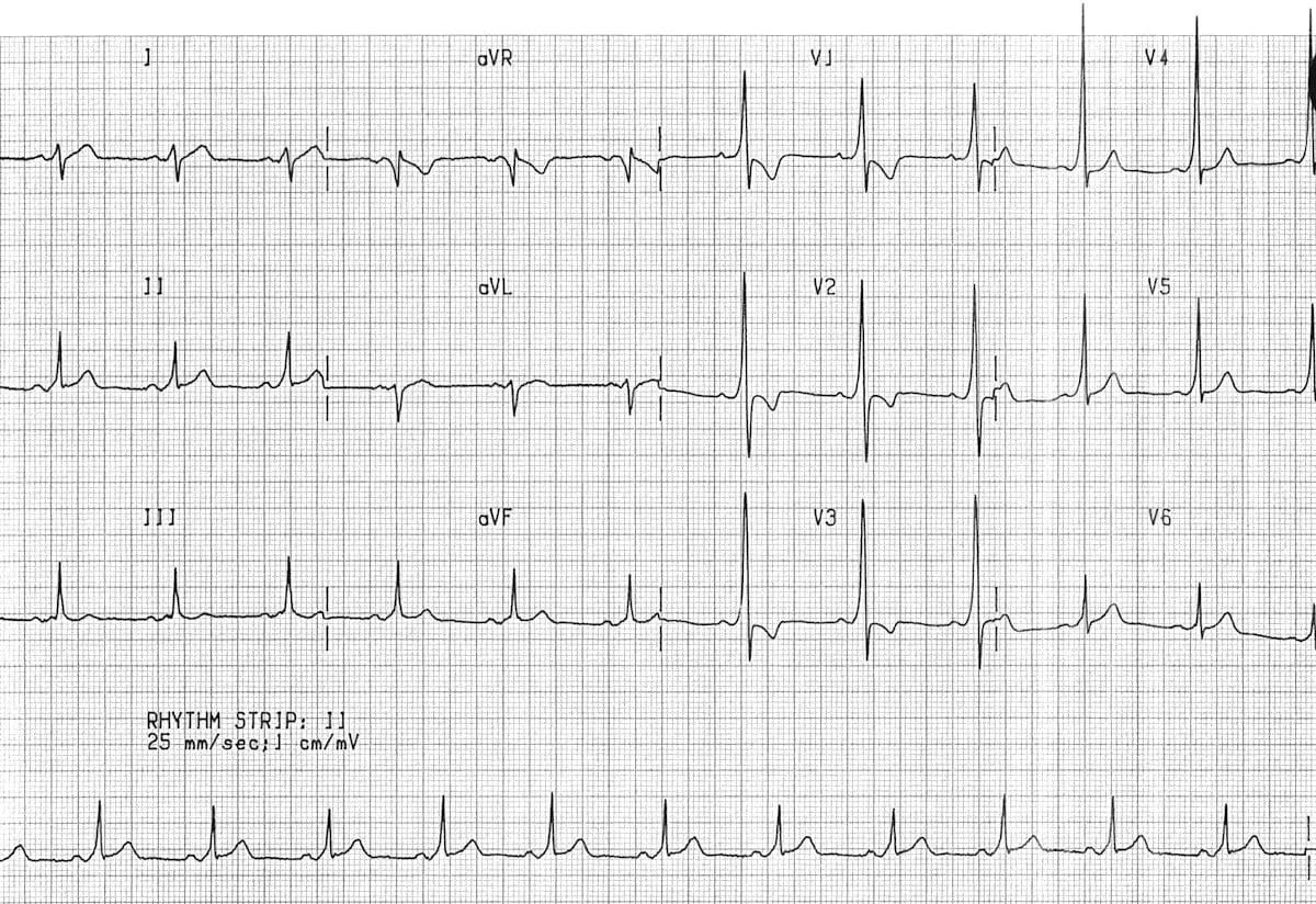
Management of PSVT
Refer to the emergency department via ambulance.
- Vagal stimulation can be attempted. Carotid sinus massage was once the treatment of choice, but has fallen out of favour given stroke risk — the Valsalva manoeuvre is generally preferred and is the best-supported of these techniques. A popular method is blowing hard into the end of an empty 20mL plastic syringe to move the plunger. Other methods: Valsalva manoeuvre (easiest for the patient), self-induced vomiting, ocular pressure (avoid — now abandoned given the risk of ocular injury), cold/ice water to the face, or swallowing ice. These manoeuvres work by stimulating the parasympathetic nervous system, slowing conduction and increasing the refractory period at the AV node.
- If vagal stimulation fails, adenosine is the first-line pharmacological agent for haemodynamically stable PSVT: it produces a brief, transient block at the AV node, interrupting the re-entry circuit. It is shorter-acting than verapamil (its effect lasts only minutes) and can be given safely to patients with heart failure or those already on a beta-blocker, unlike verapamil. IV beta-blockers, diltiazem or verapamil are alternative options for acute treatment; IV amiodarone may be considered when these are ineffective or contraindicated, and synchronised cardioversion is used if pharmacological treatment fails or is inappropriate. Adenosine (and other AV-nodal blocking agents) should be avoided if pre-excited atrial fibrillation or flutter is a possibility — see above — and any wide-complex tachycardia of uncertain origin should be managed as presumed ventricular tachycardia rather than risking a nodal-blocking agent.
Prevention and maintenance
Patients with only occasional PSVT episodes generally don’t need prophylaxis. Frequent episodes, or the presence of pre-excitation (WPW), should prompt referral to a cardiac electrophysiologist for consideration of an electrophysiological study and catheter ablation — discuss the options with the patient. Catheter ablation (radiofrequency or cryoablation) targets the slow AV-node pathway in AVNRT, or the accessory pathway itself in AVRT/WPW, and can eliminate the need for long-term medication altogether.
For prophylaxis of frequent PSVT episodes: atenolol 25mg daily, increasing if required to 100mg daily, or metoprolol. Other agents (verapamil, flecainide) may be used at a cardiologist’s discretion.