Recognition of the unwell child
Written 12th April 2020 - Last Update 22nd August 2026
Recognition of the unwell child
A structured approach: paediatric assessment triangle (PAT) → primary survey → secondary survey. See also febrile child for the age-stratified febrile workup that follows from this assessment.
Paediatric assessment triangle (PAT)
A rapid (~60 second) visual/auditory assessment before you touch the child.
- Appearance — muscle tone, mental status; reflects ventilation, oxygenation, brain perfusion and CNS/metabolic homeostasis.
- Work of breathing — body position, chest/abdominal movement, breathing pattern; abnormal sounds (grunting, wheeze, stridor); visual signs of increased work (tripoding, head bobbing, nasal flaring, gasping, tachypnoea) — reflects airway/oxygenation/ventilation adequacy.
- Circulation to skin — colour (mottling, cyanosis, pallor) — reflects cardiac output and vital-organ perfusion.
TICLS mnemonic for appearance: Tone (active vs listless) · Interactivity/mental status (alert vs disinterested) · Consolability (comforted by carer?) · Look/gaze (fixes on a face, or glassy stare?) · Speech/cry (strong, or weak/hoarse?).
Primary survey (ALS approach)
- A — obstructed or near-obstructed? Secure the airway, airway manoeuvres.
- B — work of breathing? Cyanosis? SpO₂ >94% is adequate; not breathing → assisted ventilation/CPR.
- C — pulse present? Peripherally shut down? Tachy/bradycardia? Prolonged capillary refill? Signs of hypoperfusion need IV/IO access + 20 mL/kg 0.9% saline bolus; no pulse → CPR.
- D — AVPU/GCS? Confused, floppy, seizing? Common causes found in E — hypoglycaemia, infection/fever, hypotension. Manage ABC first.
- E — look for fever, rash, BSL; expose adequately while maintaining temperature.
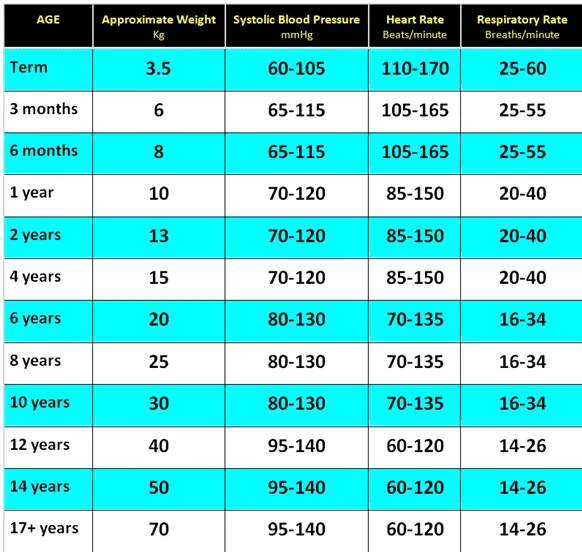
Red flags by system — neonates and young infants
Infections are the most likely cause of illness in a neonate or young infant, but several other serious conditions can look the same at first presentation, so it’s worth screening every system rather than anchoring on sepsis alone once something looks wrong:
- Breathing: grunting, tachypnoea, increased work of breathing, hypoxia, apnoea, any chest deformity.
- Circulation: pallor/mottling/cyanosis, tachy- or bradycardia, central capillary refill ≥3 seconds, a cardiac murmur, weak peripheral pulses, or an enlarged liver.
- Disability/neuro: lethargy or decreased activity, poor feeding, irritability, not responding normally to social cues, a weak/high-pitched/ continuous cry, reduced tone, or focal/prolonged seizures.
- Exposure: jaundice, a swollen limb or joint, a distended abdomen.
- Fluids/glucose: dry mucous membranes, reduced urine output, apnoea or jitteriness (both can signal hypoglycaemia).
- Infection: fever or hypothermia, a bulging fontanelle, neck stiffness, a vesicular rash (consider HSV).
A fever in any neonate (≥38°C) warrants initial investigation and empiric IV antibiotics. Unwell neonates and young infants can present with non-specific findings and may not have a fever at all — a period of observation with serial examinations is often what actually clarifies the picture, and early review by a senior clinician is essential whenever a neonate or young infant looks unwell.
Secondary survey
Complete history and detailed examination, leading to a working diagnosis and the investigations/treatment needed to confirm it.
SAMPLE history: Signs/symptoms (onset, nature, age-appropriate distress) · Allergies · Medications (exact name, dose, last dose timing) · Past medical history (immunisations, past illness) · Last food/liquid · Events leading to the injury/illness.
Assessing dehydration
Clinical signs only approximate the fluid deficit.
| Severity | Deficit | Signs |
|---|---|---|
| Mild | <4% | No clinical signs; may have increased thirst |
| Moderate | 4–6% | Delayed central capillary refill (>2 sec); increased respiratory rate; mild decreased tissue turgor |
| Severe | ≥7% | Very delayed CRT (>3 sec), mottled skin; other shock signs (tachycardia, irritability/reduced conscious level, hypotension); deep acidotic breathing; decreased tissue turgor |
The gastrointestinal tract (vomiting, diarrhoea, or both) is the most common source of significant fluid loss in children. The serum sodium at presentation changes how sick a dehydrated child looks, which is a trap worth knowing. A child with hypernatraemic dehydration tends to look more unwell for a given fluid deficit, with very dry mucous membranes and a doughy feel to the skin, but actually has relatively preserved haemodynamics — less tachycardia, better urine output — than an equally dehydrated child with a normal sodium. A dehydrated child with hyponatraemia, by contrast, may appear only mildly unwell while already being closer to hypotension and cardiovascular collapse than an equally dehydrated child with a normal or elevated sodium.
Managing the unwell child once recognised
Any neonate or young infant who appears unwell should be assessed promptly and discussed with a senior doctor. In practice this means: prompt management of suspected sepsis with early empiric IV/IM/intraosseous antibiotics (plus aciclovir if HSV is a possibility), adequate analgesia and sedation, and early referral to paediatric, surgical or sub-specialist teams as indicated. Fluid management needs to be tailored to the suspected cause, not just given at a flat maintenance rate — restrict the fluid rate to roughly two-thirds of maintenance in suspected meningitis or respiratory infection, and manage fluids judiciously where a cardiac cause is suspected. A nasogastric tube on free drainage is appropriate if bowel obstruction is suspected, and any neonate with a suspected duct-dependent congenital cardiac lesion needs early discussion with cardiology/ICU/ retrieval and consideration of IV prostaglandin to keep the ductus open.