Subconjunctival Haemorrhage
Written 18th July 2020 - Last Update 30th August 2026
Subconjunctival Haemorrhage
Blood extravasates beneath the conjunctiva, giving a sharply demarcated red patch on the sclera. It is painless, does not affect vision, and usually resolves spontaneously within 1 to 2 weeks.
Blood collects in the potential space between bulbar conjunctiva and Tenon’s capsule, so the haemorrhage stays confined and never threatens the eye itself.
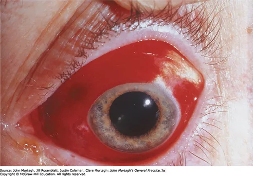
Causes
Traumatic haemorrhage is commoner in younger males — direct injury, foreign body, vigorous eye rubbing, or contact lens wear. In children, trauma accounts for most cases; one retrospective series found 83% of children with subconjunctival haemorrhage had sustained an eye injury.
Spontaneous haemorrhage is commoner in older women and becomes more frequent with advancing age, particularly after 50 years. Contributors:
- Valsalva — coughing, sneezing, vomiting, straining, heavy lifting
- Hypertension is the most consistently documented systemic risk factor, along with diabetes and dyslipidaemia
- Anticoagulants and antiplatelets, including within a therapeutic INR
- Thrombocytopenia, purpura and other bleeding diatheses
- Ocular surgery, intravitreal injection, periocular anaesthetic
- Acute haemorrhagic conjunctivitis (enterovirus 70)
Nearly half of spontaneous cases stay idiopathic.
Assessment
The diagnosis is clinical. Confirm normal visual acuity and intraocular pressure, with no pain or photophobia. Distinguish the diffuse continuous conjunctival redness from ciliary injection — a circumcorneal flush, which points to iritis, acute glaucoma, episcleritis or scleritis instead. Grittiness or a foreign-body sensation should not occur in simple subconjunctival haemorrhage.
Measure blood pressure in every patient with spontaneous or recurrent haemorrhage.
Red flags that warrant more than reassurance: recurrence, bilateral involvement, systemic bruising, or anticoagulant therapy. In those cases add a full blood count, prothrombin time, activated partial thromboplastin time and INR to look for coagulopathy or excess anticoagulation.
After trauma
Blunt trauma warrants evaluation for globe rupture and retrobulbar haematoma — both sight-threatening and needing emergency ophthalmology review. Look for proptosis, chemosis, reduced acuity or a tear-drop pupil.
If the posterior margin of the haemorrhage cannot be visualised, suspect an underlying orbital fracture — see penetrating eye injury. Haemorrhage arising from the fornix without globe trauma should prompt consideration of a basilar skull fracture. Slit lamp with fluorescein excludes a retained foreign body and corneal abrasion; orbital imaging if fracture is suspected.
In children
Suspect non-accidental injury in an infant with bilateral isolated subconjunctival haemorrhage, especially with facial petechiae. Also consider coagulopathy such as ITP or leukaemia. Newborn haemorrhage after vaginal delivery occurs in about 1-2%.
Management
No specific medical or surgical treatment is required; reassure the patient and monitor. Artificial tears help irritation but do not speed resolution. Advise avoiding eye rubbing, strenuous activity and Valsalva.
In older or anticoagulated patients, address the systemic contributors. Blood pressure control matters for recurrent or extensive bleeds. Antithrombotic therapy can generally be continued for isolated subconjunctival haemorrhage, since the ocular risk is minimal against the systemic benefit.
Recurrence happens in about 10% of cases without risk factors, and more often on anticoagulants or antiplatelets. Persistence beyond 3 weeks, or frequent recurrence, warrants reassessment for hypertension, vascular fragility or conjunctivochalasis — and any persistent conjunctival mass needs imaging or biopsy to exclude Kaposi sarcoma, melanoma or lymphoproliferative disease.