Ophthalmic Trauma (incl. Penetrating Eye Injury)
Written 17th July 2020 - Last Update 31st August 2026
Ophthalmic Trauma (incl. Penetrating Eye Injury)
Eye trauma ranges from minor abrasions to sight-threatening emergencies. AIHW data shows 86,602 Australian ED presentations for eye trauma over a 2-year period; in general practice, ophthalmic presentations (traumatic and non-traumatic) make up 1.4–1.9% of GP-trainee consultations, with trauma specifically around 0.2% of all GP visits.
History and examination
- Mechanism (blunt, penetrating, high velocity, projectile, chemical, thermal), timing, PPE worn
- Exclude associated neurological/systemic/life-threatening injury first
- Symptoms: reduced vision, redness, discharge, pain, photophobia, flashes, floaters, diplopia
- Examination: best corrected VA both eyes, IOP if available (normal 10–20 mmHg), pupil size/shape/reactivity, eye movements, visual fields, lid lacerations, subconjunctival haemorrhage/hyphaema, fluorescein staining for corneal injury
Corneal abrasion, corneal foreign body and chemical eye injury — including irrigation technique, the Roper-Hall classification and rust-ring management — now have their own dedicated page: see Corneal Abrasion, Foreign Body & Chemical Eye Injury. This page covers the more severe end of ophthalmic trauma below.
Blunt trauma
Commonly sporting (ball sports — soccer, AFL, basketball are the top three in Australian data). Key complications: traumatic hyphaema (bleeding into the anterior chamber — risk of secondary glaucoma and corneal blood staining if IOP stays elevated) and retinal dialysis (a peripheral retinal tear — an important cause of retinal detachment in younger patients).
Check VA, IOP, extraocular movements, pupil size/reactivity (traumatic mydriasis), hyphaema, and fluorescein for a Seidel-positive leak (globe rupture).
Refer if: flashes/floaters/curtain in vision (retinal injury/detachment — see
flashes-floaters.md), abnormal pupil shape/reactivity, hyphaema, loss of red reflex, high
velocity injury, or features of orbital compartment syndrome or orbital fracture (below).
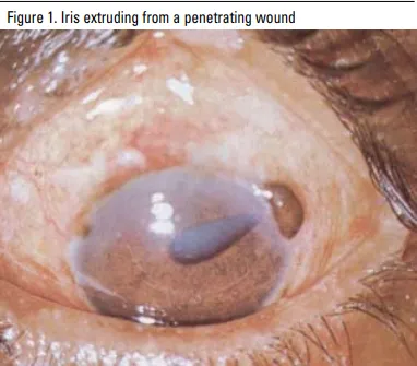
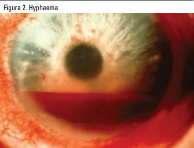
Open globe injuries (penetrating eye injury) — ophthalmic emergency
Full-thickness breach of the eye wall. Suspect with any penetrating/high-velocity mechanism, markedly reduced vision (hand movements/light perception only), a peaked/irregular pupil, or visible vitreous/uveal prolapse. The anterior chamber may look flat, with small air bubbles present; the iris may extrude from the wound in severe cases; hyphaema and lens dislocation may coexist.
Management:
- Do not touch, manipulate, or apply pressure to the eye. If an object is embedded, leave it in situ — removal risks further herniation of eye contents.
- Tape a rigid/clear shield over the eye (not a pad — e.g. the base of a polystyrene cup) — covering the eye only, not pressure-bearing
- Topical anaesthetic for comfort; antiemetics (vomiting/violent head movement can worsen extrusion); analgesia; nil by mouth
- Tetanus booster if indicated
- Advise against straining, coughing, nose-blowing or bending over before specialist review
- Urgent ophthalmology referral
Orbital compartment syndrome — time-critical emergency
Acutely raised intraorbital pressure (classically retrobulbar haemorrhage after trauma). Key signs: tight eyelids, raised IOP (often ≥40 mmHg), restricted eye movement, RAPD, proptosis, severe pain, inability to open the eye. This is a vision-threatening emergency — refer immediately to an emergency department or a centre able to perform lateral canthotomy and cantholysis (pressure-relieving procedures); delay risks permanent vision loss from optic nerve/retinal ischaemia.
Orbital fracture
Suspect with focal bony tenderness, infraorbital numbness (infraorbital nerve involvement), extraocular muscle entrapment (restricted upgaze is classic for an orbital floor “blow-out” fracture), or diplopia after blunt periorbital trauma.
Lid lacerations
Exclude intraocular injury first (slit lamp + fluorescein) and assess for concurrent systemic injury. Take clinical photographs (with consent) for the referral. Chloramphenicol ointment QID + tetanus if indicated.
Refer if: lid margin involvement, medial canthus/canalicular injury, levator injury/ ptosis, bite wounds, or any paediatric/suspected non-accidental injury.