Facial & Nasal Trauma
Written 29th June 2020 - Last Update 21st August 2026
Facial & Nasal Trauma
Primary survey
Facial trauma can accompany major trauma — assess with a standard primary survey first (Airway + C-spine, Breathing, Circulation + haemorrhage control, Disability, Exposure) before focusing on the facial injury itself.
Nasal fracture assessment
History: mechanism of injury; whether there was immediate deformity (before the onset of swelling); any new-onset nasal obstruction.
Examination: look for a septal haematoma — these need drainage, as they can obstruct the blood supply causing septal necrosis and subsequent nasal deformity, or become infected and form an abscess that can cause meningitis via venous spread.
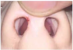
Assess nasal alignment — best done immediately, or after 48 hours to 7 days once most swelling has subsided. A nasal bone fracture causing facial deformity or obstruction needs closed reduction at 2 weeks.
Assess for CSF leak: thin, clear rhinorrhoea, usually worse leaning forward; may present from 2 days to 3 months post-injury. Refer to ED for assessment (fluid analysis for beta-2-transferrin, specific for CSF, if wanted).
Investigations
Uncomplicated nasal fractures — do not X-ray. No benefit; poorly seen; false positives and negatives. Suspected complex facial bone fractures — CT facial bones.
Management
Nasal fractures are usually managed by closed reduction under local or general anaesthesia, best done once oedema has resolved but before 2 weeks (bones start healing after this). In children, incomplete ossification means reduction is best done earlier, at 3–5 days. Per HNE practice: refer for urgent ENT assessment at 5–7 days, with closed reduction planned for 10–14 days.
Septal haematoma: refer to ED for drainage and antibiotic cover. CSF leak: refer to ED.
Anaesthesia for closed reduction: a combination of topical and local infiltrative anaesthesia (e.g. 4% cocaine or 2% lidocaine pledgets, plus 1% lidocaine with adrenaline infiltrated along the nasal bones and septum) is well tolerated in an outpatient/office setting and gives results comparable to a general anaesthetic; children, intoxicated patients, or those unable to cooperate with discomfort should have general anaesthesia instead. Closed reduction under local/topical anaesthesia has a reported success rate of 60–90% for simple, noncomminuted fractures — comminuted fractures, septal dislocation, or failure of closed reduction may need open septorhinoplasty (best delayed 3–6 months to let oedema and post-traumatic inflammation settle first). After reduction, an external dorsal splint is worn for 7 days; any internal (intranasal) splint used for septal support is removed at the same 1–2 week follow-up visit. The nasal bones remain prone to displacement for up to 2 weeks post-reduction and are not considered fully healed until 6 weeks — avoid contact sports and activities risking a blow to the nose for at least 6 weeks, and advise against nose-blowing, heavy lifting, and bending with the head below the heart in the interim.
Other facial fractures
- Zygomatic/maxillary — palpate for pain; check eye movements, assess for diplopia from restricted movement on the affected side. Usually needs discussion ± referral to maxillofacial surgery.
- Frontal sinus / orbital roof — tenderness, forehead deformity, proptosis. Assess for CSF leak (rhinorrhoea) as above. CT head/face to exclude intracranial haemorrhage. Sinus precautions for 2 weeks (avoid anything generating a Valsalva — nose-blowing, straining) to prevent orbital/intracranial surgical emphysema while the fracture heals. Maxillofacial ± neurosurgery review.
- Orbital (floor/medial wall) — periorbital haematoma, infraorbital numbness (infraorbital nerve involvement), extraocular muscle entrapment (restricted upgaze is classic for a “blow-out” fracture), or diplopia. Examine visual acuity, visual field, pupillary reflex, and eye movements. CT face to assess the size of the fracture defect; tonometry to assess intraocular pressure. Sinus precautions for 2 weeks; maxillofacial ± ophthalmology review — see Ophthalmic Trauma for the fuller orbital-fracture and orbital-compartment-syndrome work-up owned by ophthalmology.
- Mandible — presents with mandibular pain, facial asymmetry, malocclusion, trismus, or lower lip/chin numbness (inferior alveolar nerve). Examine intraorally for loose teeth, bony step-offs, and a sublingual haematoma, which is suggestive of an occult mandibular fracture even without an obvious external sign. The tongue-blade bite test is a useful bedside screen — the patient bites down and clenches a tongue depressor while the examiner twists it; an intact blade (too painful for the patient to resist) suggests a fracture, while a blade the examiner can break suggests no fracture. Bilateral mandibular body/ramus fractures can retract the anterior fragment via unopposed mylohyoid pull and compromise the airway — a red flag for urgent maxillofacial referral rather than routine review.
Facial soft tissue injury
- Treatment: tetanus booster if required; IV antibiotics if contaminated/high-risk; saline irrigation of any contaminated wound or foreign body; non-sticky wound dressing (e.g. Jelonet, gauze, Hypafix); washout and closure.
- Considerations before closure: aesthetic zones needing careful alignment (e.g. vermilion border, nasal alar rim), tissue loss, facial nerve function, and proximity/involvement of vital structures (medial/lateral canthus, parotid duct) — any of these warrant a lower threshold for maxillofacial/plastics referral rather than primary-care closure.