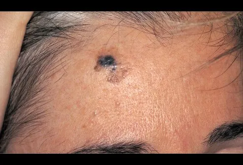
Melanoma
Written 9th April 2019 - Last Update 27th August 2026
Melanoma
Naevus classification has its own page: Naevi & Moles.
Background
Australia and New Zealand have the world’s highest melanoma rates. In Australia, melanoma is the fourth most commonly diagnosed cancer, more common in men, and the most common cancer in people aged 15–29. 1 in 14 men and 1 in 23 women are diagnosed by age 85. More common on the trunk in men, legs in women. Most present with early-stage disease (stage 1–2); 10–13% have nodal metastases (stage 3), 2–5% distant metastases (stage 4). Five-year survival: 89% (men), 94% (women) overall; 95% for thin (<1 mm), non-ulcerated, non-mitotic primaries; as low as 25% with involved nodes, thick primary, ulceration, and high mitotic count.
Risk factors and history
Any new or changing mole; personal history of melanoma/melanoma in situ; family history (risk roughly doubles per first-degree relative); sun exposure history (sunburn, protection, outdoor occupation/hobbies, tanning salons). 50–75% of melanomas arise de novo, not from a pre-existing lesion.
Examination — the ABCDEF rule
The more features present, the higher the risk:
- Asymmetry
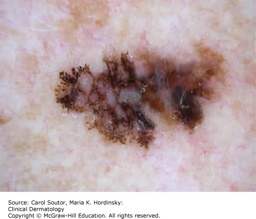
- Border irregularity
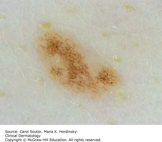
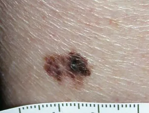
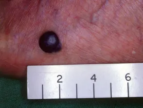
- Colour variation (several colours in one lesion; black is not essential and may be absent in nodular/amelanotic melanoma)
- Diameter >6 mm (though melanoma can be smaller)
- Evolution and/or elevation — any change in size, shape, or pigmentation
- Funny-looking — a positive “ugly duckling” sign, or a naevus that looks different to all the patient’s others
Other suspicious symptoms in a pigmented lesion: bleeding, itch, change in sensation. Perform a full skin examination (with consent, chaperone offered) including non-sun-exposed areas.
Difficult-to-diagnose forms
Melanoma is frequently misdiagnosed even by experienced clinicians:
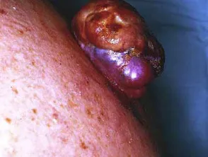
- Nodular melanoma — firm, raised, uniformly coloured, frequently non-pigmented, enlarging nodule; may bleed or become inflamed. Accounts for about 15% of melanomas, but comprises more than 60% of melanomas >3 mm in thickness. Use the EFG rule (elevated, firm, growing over 4–6 weeks) rather than ABCDE, since it often defies the classic criteria.
- Amelanotic/hypomelanotic melanoma — lacks typical pigmentation and is often not recognised as melanoma; up to 20% of all melanomas are only partially pigmented, and over a third of nodular, desmoplastic, and acral melanomas are hypomelanotic. Consider for any persistent, enlarging, or rapidly growing nodule regardless of colour — obtaining a histological diagnosis for all suspicious lesions is essential.
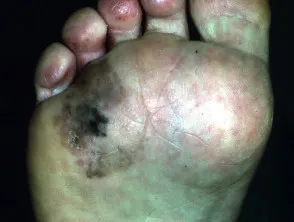
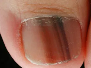
- Acral lentiginous melanoma — arises on palms, soles, fingers, toes, or beneath the nail (subungual); not sun-related; starts as an enlarging discoloured patch (often mistaken for a stain) or, in the nail matrix, an irregular pigmented longitudinal band. The most common melanoma subtype in people with darker skin. Nail apparatus melanoma specifically: rare but potentially fatal, responsible for 2–3% of all melanomas, presents as a longitudinal pigmented streak in the nail; Hutchinson sign (pigmentation of the proximal nail fold) may be present. Usually diagnosed late — mortality >50%, median survival <12 months after diagnosis, though early recognition and referral can be curative. All cases require a longitudinal nail biopsy for diagnosis; treatment is based on Breslow thickness and level of invasion (in-situ/level 1 — removal of the whole nail apparatus; invasive — amputation of the distal phalanx).
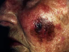
- Lentigo maligna — occurs on severely sun-damaged skin; an in-situ melanoma with a long, unpredictable delay before invasion.
- Desmoplastic melanoma — rare, aggressive, often subtle/scar-like appearance, mostly non-pigmented.
- Melanoma can also mimic seborrhoeic keratosis, SCC, or BCC.
Diagnosis
Excisional biopsy with no more than a 2.0 mm margin is preferred — it gives the most accurate Breslow thickness, ulceration, and mitotic rate assessment. Use the orientation that best facilitates a subsequent wider excision. Punch or incisional biopsy if excisional biopsy with primary closure isn’t feasible; shave biopsy is not usually appropriate but is better than missing the diagnosis. Referral without biopsy is inefficient — only consider if biopsy is genuinely problematic, and arrange dermatology or surgical referral (preferably within 7 days) for suspected primary melanoma needing biopsy beyond general-practice scope.
Managing confirmed melanoma
Margins depend on Breslow thickness (final margin also depends on other variables, not thickness alone):
- Melanoma in situ (including lentigo maligna) — wide excision with a 5 mm margin if experienced; review every 6–12 months.
- Invasive, <1 mm thick, mitotic count <2, not ulcerated, primary closure feasible — wide excision with a 10 mm margin is reasonable in general practice.
- All deeper melanomas (≥1 mm) and thin melanomas with mitotic count ≥2 — refer to a melanoma surgeon (preferably within 7 days) before any wide excision, since sentinel node biopsy assessment must precede it and cannot follow a completed wide local excision or flap repair.
Refer suspected nodal/skin/subcutaneous recurrence or metastasis (biopsy first, then refer to a melanoma surgeon within 7 days) and suspected systemic metastases (refer to a medical oncologist within 30 days).
Systemic therapy for advanced/metastatic melanoma has been transformed by immune-checkpoint inhibitors — ipilimumab (anti-CTLA-4), nivolumab/pembrolizumab (anti-PD-1), and relatlimab (anti-LAG-3), used alone or in combination. These have taken median survival for advanced melanoma from ~6 months to a ~50% long-term survival rate, and are now also used perioperatively (neoadjuvant/adjuvant) in earlier-stage disease, not just metastatic disease. BRAF tumour testing determines whether targeted BRAF/MEK inhibitor therapy is also an option. GPs should be alert to immune-related adverse events (irAEs) in patients on checkpoint inhibitors — these can affect virtually any organ system (colitis, pneumonitis, endocrinopathies, hepatitis, dermatitis) and need prompt recognition and referral back to the treating oncology team. Immunotherapy/adjuvant therapy for high-risk stage 3 disease is PBS-available.
Managing melanoma-risk conditions
- Congenital melanocytic naevi (CMN) — small/medium (<20 cm): risk does not support routine prophylactic removal; biopsy only if suspicious features develop. Large (≥20 cm): refer to a dermatologist for surveillance (baseline photography, 3-monthly review in year 1, 6-monthly for 3 years, then yearly) — prophylactic surgery has no proven melanoma-risk benefit for large/giant CMN, and lifetime surveillance is still needed regardless.
- Multiple/atypical naevi — melanoma risk rises with naevus count (>100 naevi confers 7x the risk of <15 naevi); >5 atypical naevi also raises risk. Arrange annual skin checks; excise suspicious lesions for histology.
A balancing note: melanoma overdiagnosis
The biopsy-everything-suspicious approach above remains correct practice, but GPs should be aware of a genuine, actively-debated tension in the literature: an estimated ~58% of diagnosed melanomas (22% of invasive melanomas) may represent overdiagnosis — lesions that would never have caused harm if left alone, identified using excess-incidence methodology (Welch et al.) against a background of rising diagnosis rates without a matched rise in mortality. Contributing factors include inter-pathologist diagnostic disagreement on borderline lesions and a low number-needed-to- excise threshold. This doesn’t change individual management (any concerning lesion should still be biopsied/excised, since there’s no reliable way to identify which lesions are “safe” to watch), but it’s useful context for discussing population-level skin-check programs and managing patient anxiety about excision decisions.
Follow-up and patient support
Teach the patient (and partner) self-examination and monthly self-monitoring for melanoma-in-situ patients, or 3-monthly for more significant history; 12-monthly clinician review at minimum. Primary care follow-up includes total-body checks (new primary), and suspicion for subcutaneous lumps/dermal nodules near the original site or draining nodes. Monitor vitamin D annually in winter. Provide education on recurrence risk, risk of a second primary melanoma (1 in 13 patients), and risk to first-degree relatives. Psychosocial support is often needed — body image, fear of recurrence/death, and impact on outdoor activity are common concerns.
Images