Neck Lumps
Written 9th July 2020 - Last Update 27th August 2026
Neck Lumps
Jump to: paediatric neck lumps · specific entities in more depth.
Core principle and epidemiology
Head and neck malignancy is the most common cause for neck masses in adults, and it accounts for 3.4% of all malignancies in Australia. All adult neck masses should be considered malignant until proven otherwise. Referral delay is a real, documented harm: delays in diagnosis of up to 180 days are not uncommon, with referral delay being associated with a three-fold increase in mortality.
Causes of neck swellings overall: lymph nodes (~85%), goitres (~8%), other (~7%).
Red flags and risk factors
Red flag symptoms for head and neck cancer:
- A mass present for more than two weeks.
- A recent voice change.
- Dysphagia or odynophagia.
- Ipsilateral otalgia, nasal obstruction, or epistaxis.
- Unexplained weight loss or loss of appetite.
Risk factors: smoking; alcohol use; age >40 years; a past history of head and neck malignancy or head and neck cutaneous lesions.
Additional red flag pointers: age >40 (especially >70); nodes >1.5cm (more likely to be malignant); tender mass; purple discolouration (collar-stud abscess); a simple, gradually enlarging node; fixed to skin without a punctum; associated dysphagia; hard midline thyroid lump; patient at risk of malignancy or HIV; tuberculosis exposure.
HPV-driven oropharyngeal cancer is a growing trap. The rates of human papillomavirus (HPV)-positive oropharyngeal cancer have risen markedly, with the prevalence more than tripling from 19% to 66% between 1987 and 2006. Compared with HPV-negative disease, HPV-positive head and neck cancer often do not fit the stereotype for a patient with malignancy — typically younger, male, with a higher number of sexual partners, less tobacco/alcohol exposure, marijuana use, and higher education/socioeconomic status. HPV-positive head and neck cancers are more likely to present with an asymptomatic neck mass, and the neck nodes are frequently cystic — often misdiagnosed as branchial cleft cysts, leading to delayed diagnosis and treatment.
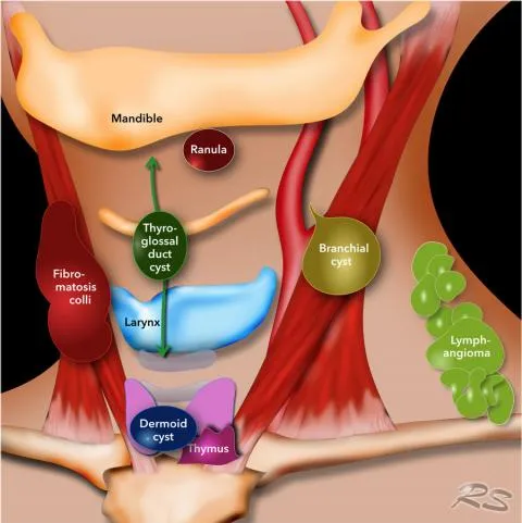
Neck lumps not due to lymph node swelling
- Widespread: sebaceous cyst; lipoma.
- Midline: thyroid nodule (moves on swallowing); thyroglossal cyst (moves on tongue protrusion — see paediatric neck lumps below for more detail); dermoid cyst (beneath the chin).
- Anterior triangle: branchial cyst (usually early adulthood — see Branchial Cleft Cyst); carotid body tumour (smooth, pulsatile, moves side to side but not vertically, age 40–60); carotid aneurysm; lateral thyroid tumours.
- Posterior triangle: developmental remnants (cystic hygroma, branchial sinuses/cysts — see below); Pancoast tumour; cervical rib.
- Submandibular: salivary gland (see below); cervicofacial actinomycosis (infection causing “lumpy jaw”).
Assessment by location — the six neck levels
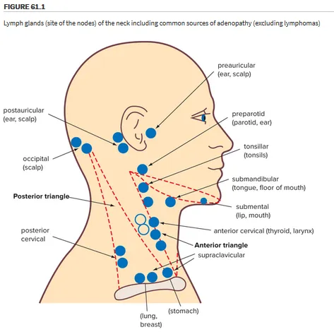
Assessing the location of a neck mass is essential as it provides clues to the aetiology. The neck is divided into six levels, each describing a lymph node region: Level I (submental and submandibular triangles); Levels II–IV (upper, middle, and lower internal jugular chain); Level V (posterior triangle); Level VI (anterior compartment).
Midline: more likely congenital — thyroglossal duct cyst is most common (can arise anywhere from the tongue base to the thyroid, most often at the hyoid level, elevates with tongue protrusion); dermoid cysts are another midline cause (fixed to overlying skin). Thyroid masses present in the midline and need evaluation given malignant potential.
Lateral: lymphadenopathy is common here (anterior triangle); branchial cysts and vascular malformations are also lateral.
Posterior triangle: higher malignancy risk. Supraclavicular lymphadenopathy is a red flag for malignancy. Consider lymphadenitis if erythema, warmth, fluctuance, induration, or severe tenderness.
Other features that need characterising: mobility of the neck mass (fixed masses are more likely to be malignant); size (>1.5cm is more likely to be malignant); firmness; overlying skin ulceration. A palpable node <1cm is normal in children; increasing size raises risk. Nodes >1cm persisting >6 weeks (or persisting despite antibiotics) should be investigated with ultrasound.
History and examination
Recent URTI, LRTI, or possible EBV, HIV, CMV, or TB exposure. Consider red flags: weight loss, dysphagia, history of cancer, increasing lump size. Palpate lymph node areas systematically, thinking about their drainage territory. Examine the lump: look, feel, move, measure, auscultate, transilluminate. Consistency is informative — hard suggests secondary carcinoma; rubbery suggests lymphoma; soft suggests sarcoidosis or infection; tender and multiple suggests infection. Palpate the midline anterior neck for thyroid lumps and the submental area for submandibular swelling.
Full head and neck examination includes inspection for cutaneous lesions, otoscopy (a unilateral middle ear effusion may indicate nasopharyngeal carcinoma), anterior rhinoscopy, oral cavity inspection and palpation, oropharyngeal inspection for masses or ulceration, tonsil enlargement or asymmetry, and flexible nasopharyngolaryngoscopy.
Investigations
FNA plus contrast-enhanced CT neck is the first-line investigation combination for a suspicious adult neck mass — together they provide complementary information on primary tumour histopathology, anatomical localisation, and nodal staging, and should be arranged simultaneously to avoid delay. Contrast-enhanced CT has an overall accuracy of 93.1% (73.3–98.0%) for FNA; for CT, studies have shown a negative predictive value of 0.33 for the detection of primary tumours and 0.57 for the detection of recurrent tumours in the head and neck — for this reason, patients with a suspicious neck mass and a normal contrast-enhanced CT should always undergo further evaluation by an ENT specialist. In indeterminate FNA cases, a repeat FNA or core biopsy may be performed — ultrasound-guided core biopsy was highly accurate (96% detection rate for malignancy) with low complication rates (1%).
Other imaging: diagnostic ultrasonography is not recommended as a first-line investigation in preference to cross-sectional imaging, being highly operator dependent, though it can guide an image-guided aspiration sample. CT is the preferred primary imaging modality over MRI (more motion artefact, longer scan times, poorer availability and tolerability), despite MRI’s better soft-tissue contrast. PET/CT has excellent sensitivity/specificity for primary and recurrent tumours but is not appropriate as a first-line imaging study given limited availability.
Ancillary tests (FBC, ANA, ESR, TSH, PTH, thyroid ultrasound, CT chest, specific infection serology for HIV/EBV/CMV/TB) assist when malignancy is unlikely or first-line investigations don’t yield a diagnosis — they should be ordered alongside, not instead of, the malignancy work-up. Basic tests used generally: FBC; ESR/CRP; CXR; TFTs (if thyroid swelling); lymph node biopsy; barium swallow if indicated.
The 20:40 rule (age-based likely cause)
- 0–20 years: congenital, inflammatory, lymphoma, tuberculosis.
- 20–40 years: inflammatory, salivary, thyroid, papillary thyroid cancer, lymphoma.
- >40 years: lymphoma, metastases — i.e. neck lumps are malignant until proven otherwise.
Management — suppurative lymphadenitis
(Systemic symptoms, erythema, tenderness, ± fluctuance.) Empirical antibiotics are widely accepted, though controversial — e.g. amoxicillin/clavulanate (duo forte) for 10 days; a 10-day course of amoxycillin/clavulanate, cephalexin or clindamycin is recognised as appropriate coverage for the most commonly involved organisms in children, and the same regimen is reasonable across ages. If symptoms have not improved with antibiotics or if the mass persists longer than four weeks, investigation with ultrasound and serological screens for atypical infections including mycobacteria… is warranted — screen for TB, Bartonella henselae/cat scratch, EBV, CMV, HIV, toxoplasmosis. Refer to a head and neck surgeon if the condition deteriorates on antibiotics.
Paediatric neck lumps
A common presentation, most often benign — congenital, inflammatory, infective, or neoplastic.
Causes. Common: thyroglossal duct cyst, reactive lymphadenopathy, lymphadenitis. Less common: branchial cleft abnormality, lymphoma, rhabdomyosarcoma, dermoid cyst, sialadenitis, malignant lymphadenopathy, salivary gland tumour, goitre/thyroid tumour, vascular or lymphatic malformation.
Reactive lymphadenopathy is by far the most common finding — 40–55% of young children have a palpable cervical lymph node as a normal finding. Cervical masses in the neonatal period and early infancy are predominantly congenital — teratoma, sternocleidomastoid tumour of infancy, vascular or lymphatic malformation. Vascular and lymphatic malformations are present at birth and typically grow commensurately with the child. A congenital mass can also first present later in childhood or adolescence, through continued growth or superimposed infection, and the likelihood of malignancy also rises in this older age group. Inflammatory pathology typically resolves within four weeks; cervical masses that persist past six weeks warrant further evaluation.
Red flags:
- Weight loss; sustained night sweats/fevers.
- Generalised lymphadenopathy; signs/symptoms of pancytopenia.
- Mass persisting >6 weeks; lymph node >3cm.
- Thyroid mass; supraclavicular mass; hard/irregular or fixed mass.
A personal history of (or exposure to someone with) an upper respiratory tract infection raises the likelihood of reactive lymphadenopathy. Animal exposure, overseas travel and tick bites are risk factors for more atypical infections such as cat-scratch disease and mycobacterial infection. A history of irradiation, or a family history of malignancy, are also malignancy risk factors worth asking about specifically.
Investigations. FBC — no strong evidence for or against routinely, but reasonable if systemic disease is suspected or infection is uncertain (may show pancytopenia or atypical cells suggesting haematological malignancy). Ultrasound is the preferred initial imaging study for most children with a neck mass — accessible, inexpensive and radiation-free, and it can guide FNA if one is needed. CT involves radiation exposure and should be avoided in children unless malignancy or a deep cervical abscess is suspected. MRI is considered the gold-standard imaging study for vascular malformations, though the frequent need for sedation limits its practical use.
FNA for diagnostic uncertainty is controversial and should only be performed by a clinician with expertise in the paediatric population. It is highly sensitive and specific… both reported up to >90%, and is less invasive than an open biopsy, but the requirement for general anaesthesia in paediatric FNA has been reported to be as high as 76%, so it’s generally a specialist-directed step.
Management. As above for suppurative lymphadenitis. Refer to a head and neck surgeon if the condition deteriorates on antibiotics, the mass persists beyond six weeks of observation, or a collection develops that needs incision and drainage. Timely referral to a head and neck surgeon for consideration of surgical management is advised for all suspected congenital neck masses, with imaging (typically ultrasound) considered within the referral process. All neck masses suspicious for a non-haematological malignancy are best referred urgently to a head and neck surgeon for further evaluation, including possible biopsy.
Specific paediatric neck masses
- Thyroglossal duct cyst — congenital, failure of thyroglossal duct closure; can present at any age (usually before adulthood); the most common midline congenital neck mass. Thyroglossal duct cysts affect approximately 7% of the population and are most commonly found in childhood, usually before ten years of age. The Sistrunk procedure (removing the central third of the hyoid bone as well) is preferred over simple excision for its lower recurrence rate. Less than 1% of thyroglossal duct cysts develop into carcinoma.
- Branchial cleft abnormality — congenital, failure of pharyngeal clefts to involute; the most common lateral congenital neck mass; can become secondarily infected. A second branchial cleft anomaly is the most common branchial cleft anomaly, with its external punctum… found anterior and medial to the sternocleidomastoid muscle. Conservative if asymptomatic, or surgical excision.
- Reactive lymphadenitis — secondary to local infection/inflammation; the most common cause of a paediatric neck mass overall; can itself become infected. Expectant management for 6 weeks; empirical antibiotics if bacterial infection suspected.
- Lymphoma — the most common malignant paediatric neck mass; rare before age 5. Hodgkin lymphoma has cervical lymphadenopathy more often than non-Hodgkin. Treated with chemotherapy/radiotherapy.
- Rhabdomyosarcoma — arises from primitive striated muscle; mostly sporadic (associated with neurofibromatosis in some cases); peak incidence age 2–5 and 15–19; the most common soft-tissue malignancy in children, presenting in the neck in ~89% of cases. Treated with surgery/chemotherapy/radiotherapy.
See also pre-auricular sinus, another common congenital head/neck lesion arising from branchial arch development.
Specific entities in more depth
Lymphatic malformation (lymphangioma)
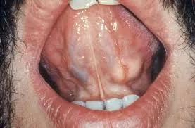
Swellings consisting of multiple lymphatic cysts made of lymphatic vessels that don’t connect to the normal lymphatic circulation. Most often found in the neck, but can occur almost anywhere. Present at birth but can subsequently grow — ranging from large fluid-filled spaces (cystic hygromas) to clusters of small firm blisters resembling frogspawn; skin-coloured, red, purple (if vascular involvement), brown, or black. Lymphangiomas are rare, accounting for around 4% of all vascular tumours and roughly a quarter of benign vascular tumours in children; there is no recognised racial or sex predilection, and most present at birth or in the first few years of life.
There are three recognised subtypes: cystic hygroma (a “macrocystic” malformation of large fluid-filled spaces, most often in the neck, axilla, or groin), cavernous lymphangioma (a rubbery skin-coloured, red, or bluish swelling that can occur anywhere on the body, including the tongue, sometimes with a phase of rapid growth in early childhood), and lymphangioma circumscriptum (a “microcystic” malformation — the frogspawn-like cluster of vesicles described above, most common on the shoulders, neck, underarm, limbs, and tongue, which can become more prominent, ooze, or bleed at puberty).
Cystic hygroma — the term historically used for very large cysts, especially in the neck, though not fundamentally different from other lymphatic malformations. Presents as a soft cystic tumour of the neck, face, or oral cavity, sometimes with visible red dots — early surgery is best; if in the mouth/pharynx, can endanger the airway. Cystic hygroma is more common in some congenital disorders, including Turner syndrome, hydrops fetalis, Down syndrome, and fetal alcohol syndrome — chromosomal analysis may be advisable in an affected infant, particularly with a posterior-neck lesion. It can now often be identified prenatally on transabdominal or transvaginal ultrasound; postnatally, ultrasound typically shows characteristic firm, rounded, non-vascular cystic spaces (distinguishing it from a venous malformation or haemangioma), with MRI reserved for more complex cases to plan treatment or define the extent of disease.
Cause: not well understood — thought to result from somatic mutations arising by chance during cell division, not inheritable, and not affected by pregnancy events. Only affects the body part where the malformation occurs.
Problems: many cause no problems at all. Most common issue is appearance; some swellings enlarge during illness (e.g. viral infection) then slowly reduce again. Pain is uncommon, sometimes from bleeding into the cyst (settles spontaneously over weeks). Infection is an uncommon but troublesome complication — can be hard to clear, and recurrent infections may permanently enlarge the malformation (longer-than-usual antibiotic courses reduce this risk). Malformations in certain locations can cause mass-effect problems (e.g. airway compromise in the neck); a large cystic hygroma involving the chin or face can also interfere with eating, breathing, or swallowing, and infection in a neck lesion can be life-threatening.
Treatment: not required if asymptomatic. Cystic hygromas and cavernous lymphangiomas are generally removed surgically, though deeper lesions can be difficult to remove completely and may regrow; injection sclerotherapy works well for large cysts, using agents such as hypertonic saline, sodium tetradecyl sulfate, doxycycline, or ethanol. Destructive options (CO2 or Nd-YAG laser, electrosurgery, cryotherapy, superficial radiotherapy) are used less often, mainly for lymphangioma circumscriptum, which is otherwise usually left untreated unless it becomes infected or symptomatic. Recurrence after surgical excision is common — reported as high as 23% for lymphangioma circumscriptum — and success is generally better for small, superficial lesions. Oral sirolimus and sildenafil are under investigation as medical treatments for severe or complicated lymphatic malformations, but carry potentially significant adverse effects and are not yet standard care. Care is needed around nerves and muscle the malformation may spread around.


Atypical mycobacterial infection
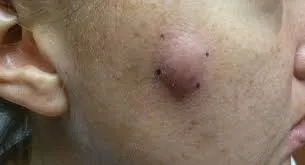
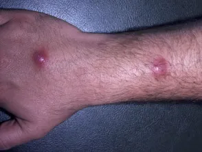
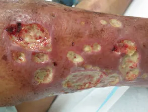
Caused by mycobacterial species other than M. tuberculosis. Can cause four clinical syndromes: pulmonary disease; lymphadenitis; skin and soft tissue disease; disseminated disease. Skin infection tends to produce crusted nodules and plaques; abscesses may develop in skin and bone infection. Each species has its own pattern. Risk factors for this and other atypical infections (e.g. cat-scratch disease) affecting the neck include animal exposure, overseas travel, and tick bites (see paediatric red flags above) — worth asking about specifically when a neck lump has failed to respond to standard empirical antibiotics.
Cervicofacial lymphadenitis — the pattern most often seen in general practice — is caused by Mycobacterium avium complex in around 80% of paediatric cases. It typically affects children under 5, usually as a single soft but firm, non-tender, submandibular or cervical-chain mass that slowly enlarges over days to months; later stages develop tenderness and necrosis within the node, then overlying erythema, then skin breakdown with sinus tract formation. The diagnosis is frequently suspected only after enlarged cervical nodes fail to respond to empirical antibiotics aimed at Staphylococcus/Streptococcus, or fail to resolve on watchful waiting for a presumed viral cause.
Treatment depends on the organism isolated — standard TB treatment doesn’t work for most of them; rifampicin is a common effective agent across most species. For cervicofacial lymphadenitis, a macrolide (azithromycin or clarithromycin) combined with rifampin or ethambutol is the typical antibiotic regimen, continued until symptoms resolve; combining antibiotics with surgical excision of the infected node significantly increases cure rates (around 95%, versus roughly two-thirds with antibiotics alone), though excision of preauricular nodes carries a risk of facial nerve injury as the nerve courses through the underlying parotid gland — a risk worth weighing against the somewhat lower cure rate of antibiotics alone, especially in young children.
Salivary gland tumours
85% occur in the parotid gland, then submandibular and minor salivary glands, with only 1% in the sublingual glands. 80% are benign — slow-growing, movable, painless, usually solitary, found beneath normal skin or mucosa; some are cystic (soft) but most are firm. Confusingly, the risk of malignancy runs the opposite way to the frequency of tumours — it is higher in the smaller glands (minor salivary and sublingual) than in the parotid, even though the parotid is where most tumours of any kind occur.
Risk factors: older age; radiation exposure (e.g. prior head/neck cancer treatment); workplace exposure (rubber manufacturing, asbestos mining, plumbing).
Symptoms:
- A lump/swelling near the jaw, neck, or mouth.
- Facial numbness; unilateral facial muscle weakness.
- Persistent salivary gland pain; dysphagia; trouble opening the mouth widely.
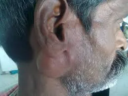
When examining a parotid swelling, distinguish a focal, discrete mass (which may be a tumour, an enlarged intraparotid lymph node, or an abscess, and warrants imaging) from diffuse swelling (more typical of parotitis). Formal assessment of facial nerve (CN VII) function is essential in any parotid mass, since CN VII branch palsy is commonly associated with parotid malignancy — though it is occasionally also seen with benign infective parotitis or a parotid abscess, so an urgent ENT referral to exclude malignancy is warranted either way.
Benign: the most common is pleomorphic adenoma (mixed tumour) — can transform malignantly, but usually only after 15–20 years; if malignant transformation occurs, the cure rate is very low, so surgical treatment is often recommended even at the benign stage. Other benign tumours (monomorphic adenoma, oncocytoma, Warthin tumour) rarely recur or transform.
Malignant: less common, characterised by rapid/sudden growth spurts; firm, nodular, may be fixed to adjacent tissue; overlying tissue may ulcerate or adjacent tissue may be invaded.
- Mucoepidermoid carcinoma — the most common salivary gland cancer; age 20–50; most commonly parotid; intermediate/high-grade tumours can metastasise to regional lymphatics. Treatment is wide excision with postoperative radiation for high-grade lesions; the 5-year survival rate is over 90% for the low-grade type but under 50% for the high-grade type.
- Adenoid cystic carcinoma — the most common malignant tumour of the minor salivary glands (and trachea); slow-growing, peak incidence age 40–60; concerning for severe pain and facial nerve paralysis given its tendency to invade and spread along perineural tissue, sometimes many centimetres from the original tumour. Lymphatic spread is uncommon, but late distant metastasis (usually pulmonary) is common, and cause-specific survival keeps declining for up to 30 years after diagnosis despite favourable 5- and 10-year survival figures.
- Acinic cell carcinoma — a common parotid tumour, age 40s–50s, more indolent course, can be multifocal.
- Carcinoma ex mixed tumour — adenocarcinoma arising in a pre-existing benign mixed tumour; only the carcinomatous element metastasises. Primary treatment is parotidectomy aiming for complete resection, with neck dissection if there is nodal disease (and considered even without evidence of nodal spread in some patients).
All surgery for salivary gland malignancy is designed to spare the facial nerve, which is sacrificed only if directly involved by tumour.
Diagnosis: FNA biopsy for tissue diagnosis; CT and MRI for extent of disease. Treatment: surgery, sometimes with radiation, for malignant disease.