Vulvar disorders
Written 23rd April 2020 - Last Update 27th August 2026
Vulvar disorders
Vulvar discomfort/irritation: diagnostic strategy model
Serious disorders not to be missed: neoplasia (vulval intraepithelial neoplasia, squamous cell carcinoma, Paget’s disease, melanoma), provoked vestibulodynia.
Pitfalls (often missed): psoriasis, lichen planus, lichen sclerosus, lichen simplex chronicus, aphthous ulcers, dysaesthetic vulvodynia, trichomonal vaginitis, herpes simplex virus, infestations (threadworms, pubic lice, scabies).
Seven masquerades: depression, diabetes, drugs, spinal dysfunction, UTI.
Is the patient trying to tell me something: commonly, psychosexual problems.
Causes of common vulval signs
- White patches: lichen simplex chronicus (lichenification), lichen sclerosus, vitiligo, vulval intraepithelial neoplasia.
- Erosions and ulcers: herpes simplex or zoster, lichen planus, aphthous ulcers, desquamative inflammatory vulvovaginitis, contact dermatitis, fixed drug reaction, excoriated scabies, Crohn’s disease of the vulva.
- Intertrigo (inflammation of skin folds): atopic dermatitis, Candida albicans, seborrhoeic dermatitis, tinea, erythrasma, psoriasis.
Vulval dermatitis
Very common. Think about and ask around: atopic dermatitis, irritant contact dermatitis, allergic contact dermatitis, seborrhoeic dermatitis, corticosteroid-induced dermatitis, psoriasis.
- Examination: expect some inflammation; look carefully for lichenification.
- Investigations: reasonable to take a vaginal swab for candida; cervical screening test if due; consider vulval biopsy if a malignant condition is suspected, especially with thickening or textural change.
- Management — vulval care: avoid scented/dyed products (toilet paper, washing powder, lotions, deodorants), tight/synthetic/uncomfortable clothing, abrasive activities (cycling, horse riding), daily panty liners or pads, and vaginal douches, wipes or deodorants. Use an aqueous cream or warm-water soaks instead of soap, and a bland emollient such as white soft paraffin. 1% hydrocortisone ointment twice daily for 4 weeks; a stronger steroid may be needed if there’s an endogenous skin condition such as eczema or psoriasis.
Lichen sclerosus
Unclear cause, classified as an autoimmune disease.
-
Demographics: most commonly diagnosed in women over 50; can occur in prepubertal girls.
-
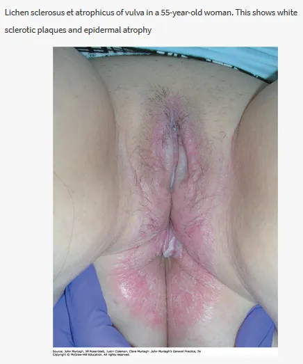
Murtagh’s triad: genital pruritus + soreness + white wrinkled plaques → lichen sclerosus.
-
History: 15% have a family member with lichen sclerosus; often associated with other autoimmune disease (e.g. thyroid disease, pernicious anaemia, alopecia areata); often extremely itchy and sore.
-
Examination: primarily involves the non-hair-bearing, inner areas of the vulva — can be localised to one small area or extensively involve the perineum, labia minora and clitoral hood; can spread onto the surrounding labia majora and inguinal fold, and in 50% of women to the anal and perianal skin; never involves the vaginal mucosa. Can cause adhesions and scarring — the clitoris may be buried, the labia minora resorb/shrink, and the vaginal entrance tightens (introital stenosis).
-
Investigations: skin biopsy is recommended.
-
Management: refer to a specialist dermatologist or gynaecologist. Development of squamous cell carcinoma occurs in 4–5%. Ultrapotent topical steroids are often prescribed.
Lichen planus
Can affect the vulva and vagina. Autoimmune disorder.
- History: painful and persistent erosions; painful desquamative vaginitis preventing intercourse and causing a mucky vaginal discharge — the eroded vagina may bleed easily on contact; overlaps with lichen sclerosus. Contributing factors: genetic predisposition, physical and emotional stress, injury to the skin, localised skin disease such as herpes zoster, systemic viral infection such as hepatitis C, and drugs (gold, quinine, quinidine often cause a lichenoid rash).
- Examination: may affect the labia majora, labia minora and vaginal introitus — painless white streaks in a lacy or fern-like pattern; scarring resulting in adhesions, resorption of the labia minora and introital stenosis.
- Investigations: often requires a biopsy to confirm diagnosis.
- Management: refer to a specialist to rule out lichen sclerosus. Treatment isn’t always necessary but can include potent topical steroids, tacrolimus or pimecrolimus cream, topical retinoids, or intralesional steroid injections. Can rarely progress to squamous cell carcinoma.
Lichen sclerosus — practical points (AJGP)
Common (~1 in 70 women), all ages but most often postmenopausal, and typically diagnosed 5–15 years late because of patient embarrassment and clinician under-recognition.
- Distribution: white plaques, hyperkeratosis and purpura in a figure-of-eight around the vulva and perianal skin, never involving the vaginal mucosa. Architectural change (labial resorption, buried clitoris, midline fusion, introital narrowing) marks chronicity; pallor and purpura/fissuring mark active disease.
- Treatment: an ultrapotent topical corticosteroid ointment (not cream — better penetration, barrier function and lower allergen risk), usually clobetasol propionate 0.05% (now PBS-listed as Xobet, previously only available compounded). Australian consensus favours lifelong suppression, not on-demand use for symptoms. A typical induction “blitz” is daily for 1 month, alternate days for 1 month, then 2–3 times/week maintenance — not a one-off course. Apply to the affected vulva only, in a thin layer (a half-pea-sized quantity), avoiding hair-bearing skin (misapplication to the labia majora causes steroid telangiectasia/steroid rosacea). Skin biopsy where the diagnosis is uncertain or to exclude neoplasia.
- Stable disease still needs lifelong 12-monthly examination (symptom control alone is not a reliable indicator of adequate treatment) — assessing symptoms, disease activity, architectural change and malignancy. Clinical photography is the gold standard for tracking architectural change over time; patient self-examination (with a hand mirror) is a useful adjunct once disease is stable.
- Malignancy surveillance: vulval SCC arising on a background of LS is HPV-independent (~2.2% risk) via a differentiated VIN (dVIN) precursor (~1.2% risk) — distinct from the HPV-dependent pathway (HSIL/usual-type VIN) seen without LS, and associated with worse treatment resistance, recurrence and mortality. Adequate topical corticosteroid therapy significantly reduces this malignancy risk — review long-term, and biopsy any non-healing ulcer, erosion, thickening or fixed lump.
- Steroid phobia is a major, underappreciated barrier to adherence — proactively counter it with clear, consistent counselling (multiple cohort studies show long-term topical corticosteroid use in LS has no irreversible local or systemic adverse effects).
- Sexual dysfunction affects up to 50% (dyspareunia, reduced orgasm quality, distress about genital self-image) — consider psychosexual counselling and pelvic floor physiotherapy. LS is also associated with depression (40–58%) and anxiety (~27%) in published cohorts, and with autoimmune comorbidities (thyroid disease, pernicious anaemia, vitiligo, psoriasis) — screen and manage accordingly.
- Persistent or worsening vulval itch without objective LS activity on exam should prompt reassessment for a coexisting/alternative cause — vulvovaginal candidiasis, genitourinary syndrome of menopause, contact dermatitis, eczema or psoriasis — rather than assuming it reflects LS activity.
A vulvar ulcer that does not heal is cancer until proven otherwise. A persistent ulcer — especially in a postmenopausal woman or on a background of lichen sclerosus — should not be treated presumptively as infection indefinitely; biopsy to exclude squamous cell carcinoma / differentiated VIN.
Bartholin’s cyst and abscess
The Bartholin’s (greater vestibular) glands sit at the 4 and 8 o’clock positions of the vaginal introitus and secrete lubricating mucus. Duct obstruction → a Bartholin’s cyst; secondary infection → a Bartholin’s abscess.
- Cyst: usually a painless, unilateral, fluctuant swelling of the posterior labium; often incidental. Small asymptomatic cysts need no treatment.
- Abscess: rapidly enlarging, exquisitely tender, red, hot unilateral labial swelling ± difficulty sitting/walking; may point and discharge. Often polymicrobial (gut/skin flora); consider gonorrhoea/chlamydia and take swabs.
- Management: simple incision and drainage alone has a high recurrence rate — the definitive procedure is Word catheter insertion (a small balloon catheter left in for ~4 weeks to epithelialise a drainage tract) or marsupialisation. Antibiotics are added if surrounding cellulitis, systemic features, pregnancy, or immunosuppression.
- New or solid Bartholin’s mass in a woman >40 → refer to exclude the rare Bartholin’s gland carcinoma (biopsy/excision rather than simple drainage).