Ovarian Cysts, Masses & Accidents
Written 20th April 2020 - Last Update 18th July 2026
Ovarian Cysts, Masses & Accidents
Assessment of an adnexal cyst/mass — including malignancy risk stratification with the Risk of Malignancy Index — and the two acute “ovarian accidents” (cyst rupture and torsion). Ovarian malignancy itself is on Ovarian cancer; this page is the benign/undifferentiated cyst work-up plus the acute presentations.
Types of ovarian cyst/mass
- Functional (follicular or corpus luteum) — the commonest, physiological.
- Endometrioma (endometriosis deposits within the ovary).
- Cystadenoma (serous or mucinous), dermoid (mature teratoma — hair, skin, teeth, fat), borderline tumour, ovarian cancer.
Non-ovarian masses that mimic a cyst on imaging: paratubal/paraovarian cyst, hydrosalpinx, appendiceal or retroperitoneal cyst, pedunculated fibroid.
Assessment
Usually an incidental finding on examination or ultrasound; may cause lower abdominal/pelvic pain. Always screen for ovarian-cancer symptoms (persistent abdominal distension/increased girth, early satiety, pelvic pain, new urinary frequency/urgency).
- ≥2 of these symptoms persisting >2 weeks → pelvic ultrasound (transabdominal + transvaginal) ± CA-125 to exclude ovarian cancer (particularly older women).
- Cyst found on ultrasound:
- CA-125 for all postmenopausal patients.
- CA-125 + AFP, β-hCG, LDH for premenopausal patients with complex cysts (germ-cell tumour markers).
Risk of Malignancy Index (RMI)
RMI = ultrasound score × menopausal score × CA-125 (U/mL).
| Component | Score |
|---|---|
| Ultrasound features (multilocular cyst, solid area, metastases, ascites, bilateral) | 0 = none · 1 = one feature · 3 = ≥2 features |
| Menopausal status | 1 = premenopausal · 3 = postmenopausal |
| CA-125 | actual value (U/mL) |
- RMI 25–200 — moderate risk → gynaecologist.
- RMI >200 — high risk → gynaecological oncologist.
- RMI can be falsely normal in malignancy — refer to gynae-oncology if RMI = 0 but CA-125 >80 U/mL, or if clinically suspicious.
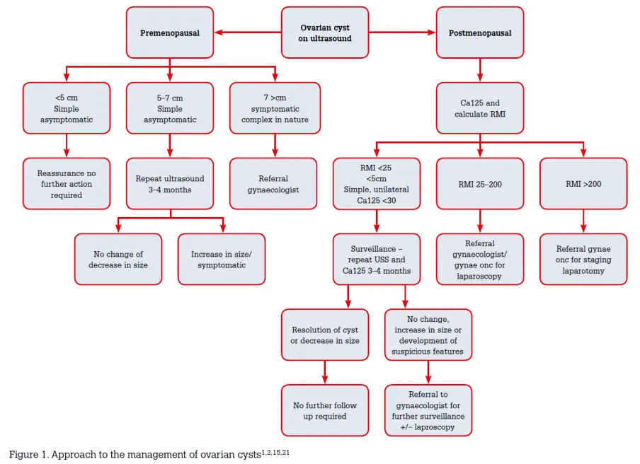
Management of a cyst by age
A 3–4 cm simple cyst is normal in a woman of reproductive age.
Children/adolescents (≤15): any cyst, any size/type → urgent gynaecology assessment.
Premenopausal (almost always benign; malignancy ~1/1000, up to ~3/1000 by 50):
- Simple cyst <6 cm asymptomatic → repeat ultrasound in 4–6 months.
- Simple cyst <6 cm with significant (ovarian-cancer-type) symptoms → routine gynaecology (may be incidental).
- Simple cyst ≥6 cm → seek gynaecology advice.
- Complex cyst: tumour markers raised → gynae-oncology; normal → repeat ultrasound at 6 weeks (persistent → routine gynae; enlarging → gynae-oncology).
- Dermoid, cystadenoma, or endometrioma identified → routine gynaecology.
Postmenopausal (a cyst here is abnormal — always refer):
- Simple, unilateral, unilocular, <5 cm, RMI 25–200 → routine gynaecology.
-
5 cm, complex features, RMI >200, or CA-125 >80 → gynae-oncology.
Acute ovarian accidents
Ovarian cyst rupture
Physiological rupture of a small follicular cyst at ovulation is usually asymptomatic or causes mild mid-cycle pain (mittelschmerz). Symptomatic rupture releases fluid/blood that irritates the peritoneum.
- Presentation: sudden severe focal unilateral lower-quadrant pain, often during exercise or intercourse; right ovary most commonly (the rectosigmoid protects the left). Massive haemorrhage → shoulder-tip/upper-abdominal pain (subphrenic blood), ± Cullen’s sign. Dermoid rupture causes severe chemical peritonitis and usually needs surgery.
- Risk: ovulation induction increases risk; oestrogen-progestogen contraception reduces it.
- Investigations: pregnancy test (exclude ectopic), FBC (Hb may be normal early), group & crossmatch if peritoneal signs/instability, pelvic ultrasound (adnexal mass + free fluid in the pouch of Douglas).
- Management: uncomplicated (physiological) rupture → oral analgesia; fluid resorbs within ~24 h, symptoms settle over a few days. Heavy/ongoing bleeding → inpatient observation ± laparoscopy; in a premenopausal woman, ovarian-sparing cystectomy is preferred over oophorectomy.
Ovarian (adnexal) torsion
Total/partial rotation of the ovary around its vascular pedicle — a surgical emergency. Early venous/lymphatic obstruction (with continued arterial inflow) swells the ovary; untreated, arterial occlusion causes haemorrhagic infarction and necrosis.
- Right-sided in ~⅔ (the sigmoid limits left-ovary mobility). Adnexal torsion usually involves both ovary and tube.
- Risk factors: an ovarian mass (present in >half — usually benign, dermoid most common; malignant tumours torse less, being fixed by adhesions), pregnancy (~5×, greatest in the first trimester), assisted conception (enlarged ovaries), previous pelvic/tubal surgery, developmental abnormalities.
- Presentation: sudden severe unilateral lower-abdominal pain that worsens intermittently, with nausea/vomiting; a tender adnexal mass. Late necrosis → fever, tachycardia, hypotension, peritonism. Hard to distinguish from appendicitis on the right.
- Investigations: primarily a clinical diagnosis. β-hCG (exclude pregnancy); bloods are non-specific. Doppler ultrasound may show an enlarged ovary, peripheral follicles, a midline ovary, free fluid, absent parenchymal perfusion, or the “whirlpool sign” — but normal Doppler flow does not exclude torsion.
- Management: urgent ED/gynaecology referral for laparoscopic detorsion ± oophoropexy; salpingo-oophorectomy only if the ovary is frankly necrotic or peritonitis is established.