Breast Cancer & Screening
Written 20th April 2020 - Last Update 12th October 2026
Breast Cancer & Screening
Covers breast malignancy, population screening and risk-based prevention, and Paget disease of the nipple. The assessment of a symptomatic breast (lump, pain, discharge) is on Breast symptoms — any symptomatic lump is worked up with the triple test, not screening mammography.
Breast cancer
Histology: ~90% invasive ductal carcinoma; the rest lobular, papillary, medullary, mucoid/colloid. Ductal carcinoma in situ (DCIS) is a non-invasive precursor (~20% of breast cancers), treated with excision (lumpectomy vs mastectomy) ± radiotherapy, ~98% survival.
Epidemiology: incidence rises with age — uncommon before 30, peak ~60; roughly one third pre-menopausal, two thirds post-menopausal. Lifetime risk ~1 in 8 (13%) by age 85. More than 75% of cases occur in women over 50.
Risk factors
- Age (the strongest non-genetic factor — age 50 carries ~10× the risk of age 30).
- Genetic: BRCA1 (RR ~5.9) and BRCA2 (RR ~3.3); also PALB2, PTEN, CHEK2, ATM, CDH1 (lobular), STK11; polygenic risk scores. At least ~5% of breast cancers are due to inherited high-penetrance variants. Ashkenazi Jewish ancestry.
- Family history (first-degree relative ~3-fold), personal history of breast cancer (~1%/year recurrence), previous chest/breast radiation.
- Oestrogen exposure: early menarche (<12), late menopause (>53), nulliparity, first child after 30, prolonged MHT (>5 years).
- Proliferative benign disease (atypical hyperplasia, LCIS), high mammographic density.
- Modifiable: alcohol (>2 standard drinks/day), elevated BMI, physical inactivity.
Presentation
- Painless hard, irregular lump — ~76% present with a lump; pain is the presenting feature in only ~10%.
- Nipple change: discharge, retraction, distortion; Paget disease (below).
- Skin dimpling/puckering, oedema (peau d’orange).
- Rarely, presents with bony secondaries (bone pain, weight loss).
Investigation & management (GP level)
- Every palpable symptomatic lump gets a triple test (see Breast symptoms) — an abnormality at any stage prompts breast-surgeon referral.
- Prompt referral to a breast surgeon for anything suspicious.
- Accurate staging requires lymph-node assessment (sentinel node biopsy) — nodal status is the best predictor of distant spread. Definitive treatment combines surgery, radiotherapy, chemotherapy and endocrine/targeted therapy.
- Specialist treatment has expanded well beyond the note’s original era — CDK4/6 inhibitors for HR-positive disease, immunotherapy and antibody–drug conjugates for triple-negative disease — but this does not change the GP assessment and referral pathway above.
Screening (BreastScreen Australia & the Red Book)
- Biennial (2-yearly) mammography, ages 50–74, is the actively invited program. Ages 40–49 and 75+ are eligible but not actively invited.
- Aboriginal and Torres Strait Islander women are recommended to screen from age 40, earlier than the general 50-year invitation age, given lower participation and later-stage presentation.
- Screening in those >75 is reasonable if life expectancy is >10 years.
- The 60–69 age band has the most favourable benefit; evidence in 40–49 is least favourable.
- Trans and gender-diverse people: screen by anatomy. Trans men without chest surgery screen as usual. For trans women, most states recommend 2-yearly screening at 50–74 after at least 5 years on GAHT; WA does not. Explain that the screening service may ask about GAHT because it increases breast density. See Gender Dysphoria.
Screening is imperfect. Biennial mammography reduces breast-cancer mortality by ~15–20% but with meaningful overdiagnosis (older trial estimates ~30% of screen-detected cancers). Counsel women that screening detects, but does not prevent, cancer.
Breast density
Density is now notified. BreastScreen Australia’s 2024 position statement made it national policy that women are informed in writing of their mammographic density on their screening mammogram.
Increased breast density reduces the sensitivity of screening mammography, and modestly increases the risk of breast cancer.
| BI-RADS | Description | Sensitivity | Risk vs BI-RADS B |
|---|---|---|---|
| A | Almost entirely fatty | ~90% | ~0.5× |
| B | Scattered fibroglandular density | ~90% | 1 (reference) |
| C | Heterogeneously dense | ~84% | 1.6× |
| D | Extremely dense | ~64% | 2.6× |
Dense breasts” = BI-RADS C or D. “About 33% of women aged 50–74 are C or D, and about 10% are D (rising to ~25% of women under 50). Density generally falls with age.
Despite the lower sensitivity, mammography remains the screening test for a population program at every density.
Breast Density In Clinical Practice
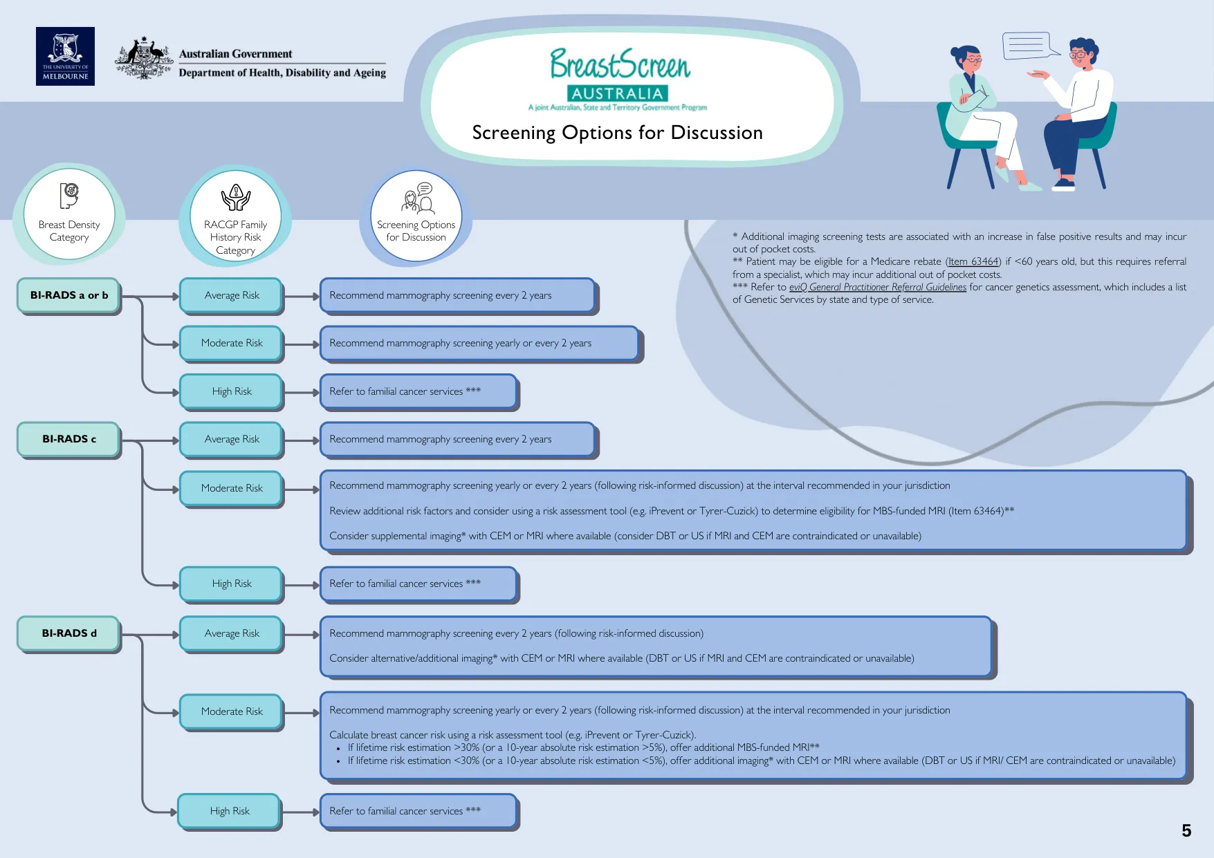
The pathway depends on density category & risk status — not on density alone:
- Moderate family-history risk, at any density — yearly or 2-yearly (at the interval recommended in your jurisdiction, after a risk-informed discussion), rather than routine biennial screening.
- BI-RADS D + average risk — still 2-yearly mammography, but supplemental imaging should be discussed (CEM or MRI where available; DBT or US if those are unavailable or contraindicated). This is the group most likely to be missed, because average family-history risk otherwise reads as “nothing to do here”.
- BI-RADS C or D + moderate risk — calculate absolute risk with a validated tool (below) to test MBS-funded MRI eligibility, then consider supplemental imaging.
- High family-history risk, at any density — refer to a familial cancer service per the eviQ GP referral guidelines. Density does not change this and does not need to be resolved first.
- BI-RADS A/B or C at average risk — unchanged, routine 2-yearly mammography.
Supplemental imaging
There is still no consensus on the optimal supplemental test, and no RCT showing supplemental screening saves lives in asymptomatic women without other risk factors — the DENSE trial showed MRI finds more cancers in extremely dense breasts, but did not directly measure mortality benefit. All supplemental imaging increases false positives, callbacks, benign biopsies, anxiety and cost.
| Test | Extra cancers /1000 screened | Trade-offs | MBS |
|---|---|---|---|
| MRI | ~16 | Access, cost, false positives, claustrophobia, gadolinium | Item 63464 — high risk by risk tool, age <60, specialist referral required |
| CEM | ~16 | Access, cost, false positives, radiation, iodinated contrast | No item for supplemental screening |
| Ultrasound | ~4 | Widely available, cheap, operator-dependent, highest false positives | Item 55076 if mammography inconclusive due to density |
| DBT | ~1.7 | Widely available, cheap, extra radiation | Item 59302 — significant family history / clinical indication |
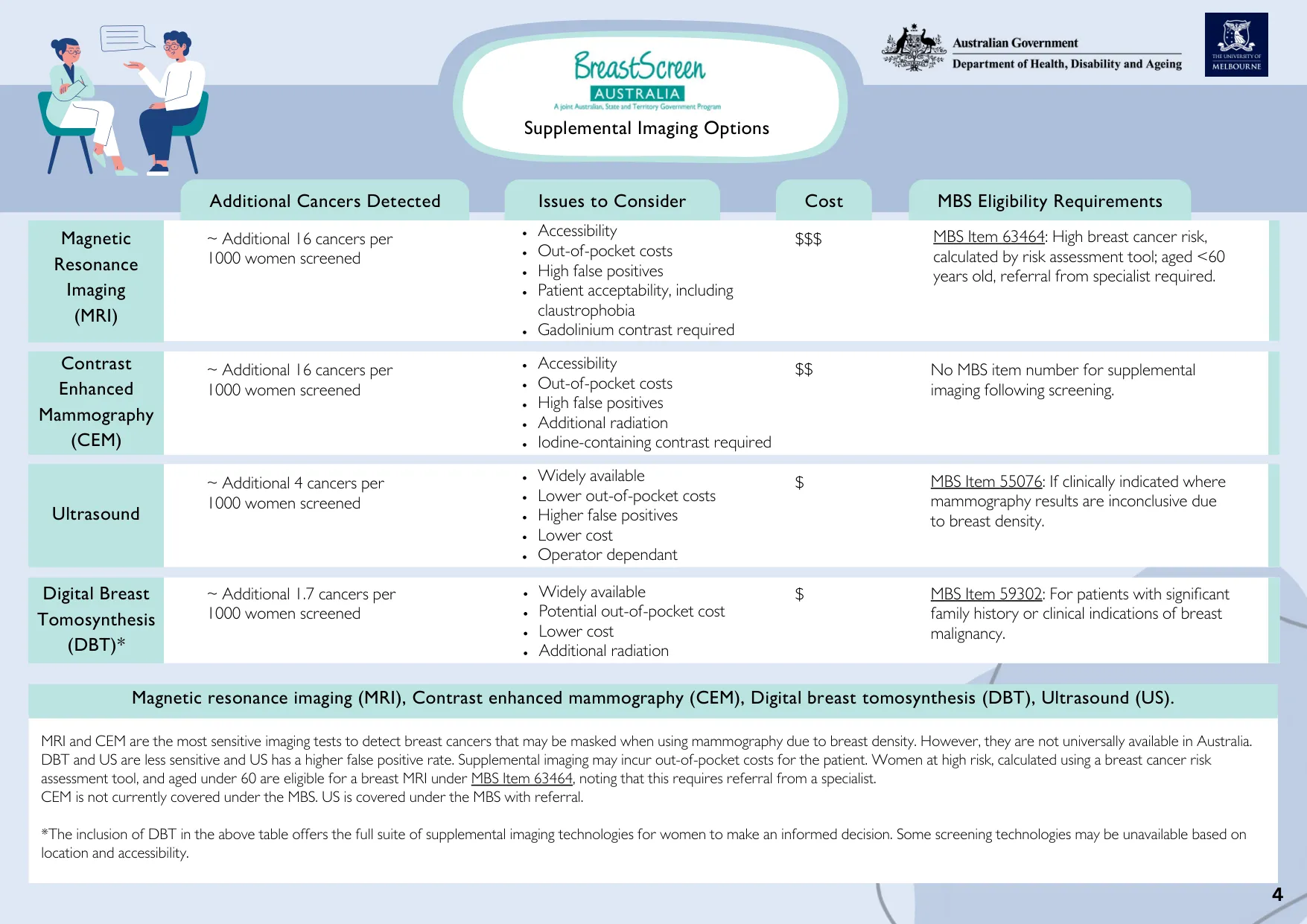
MRI and CEM are the most sensitive for cancers masked by density, but are not universally available in Australia. The practical trap is MBS item 63464: it needs a calculated high risk and age under 60 and a specialist referral — a GP cannot generate the rebate directly. Women who don’t qualify are looking at self-funded CEM or MRI.
Both figures above are reproduced from the BreastScreen Australia Breast Density GP Guidance (November 2025) — see the Sources page.
Risk assessment
Use a validated tool (iPrevent, Tyrer-Cuzick, FRA-BOC/CanRisk) and reassess periodically. These tools combine family history, genetic risk, hormonal and lifestyle factors — and iPrevent and Tyrer-Cuzick both incorporate breast density, which is why they are the ones named in the density pathway above.
iPrevent (Peter MacCallum Cancer Centre) is the usual Australian choice — free, online, validated, and it produces both a risk estimate and a management-options output to work through with the patient.
The number that matters for access is the MBS MRI threshold: lifetime risk >30%, or 10-year absolute risk >5% (item 63464, age <60, specialist referral). Below that threshold, supplemental imaging is self-funded.
The FRA-BOC risk categories:
- Category 1 — average / slightly above average (~1–1.5× population, ~95% of women; ~9–12.5% risk to 75): normal screening schedule.
- Category 2 — moderately increased (~12.5–25% risk; e.g. one first-degree relative with breast cancer <50): annual mammography from 40.
- Category 3 — potentially high risk (~25–50%; e.g. ≥2 first/second-degree relatives plus high-risk features — diagnosis <45, Ashkenazi ancestry, male relative with breast cancer): full FRA-BOC assessment and referral to a family cancer clinic for genetic testing and an individualised surveillance plan (which may include MRI).
Prevention
- Breast awareness — familiarity with the normal look/feel of one’s breasts and prompt reporting of new change. No single self-examination technique is superior; routine clinical breast examination is not helpful for average/moderate-risk women (may have a role in high-risk surveillance).
- Lifestyle: limit alcohol, maintain a healthy weight, physical activity.
- Risk-reducing medication (tamoxifen, raloxifene, or an aromatase inhibitor) and risk-reducing surgery can be offered to women at moderate-to-high risk — see the COSA 2024 patient guide; refer for specialist counselling.
Paget disease of the nipple
A sign, not a diagnosis — a marker of underlying breast malignancy.
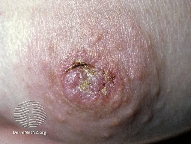
- Starts as a red, dry, scaling, eczematous rash of the nipple and areola that progresses to ulceration. It begins centrally on the nipple and spreads out to the areola and then the surrounding skin. It can look identical to eczema — patients are typically treated with topical steroids for months before referral.
- Any eczematous nipple/areolar change not resolving after 2 weeks of topical therapy → refer to a breast surgeon and assume cancer. Do not wait if there are other red flags (lump, nipple distortion, discharge).
Tamoxifen and the endometrium
Tamoxifen (a SERM — anti-oestrogenic in breast, oestrogenic in the endometrium) is standard adjuvant endocrine therapy for oestrogen-receptor-positive disease. GP-relevant points:
- No routine endometrial screening (ultrasound or biopsy) in asymptomatic women on tamoxifen — endometrial-cancer incidence is low (2–3/1000 women/year).
- Any abnormal bleeding needs prompt investigation with a low threshold for biopsy — cumulative endometrial-cancer risk is ~1.6% at 5 years, ~3.1% with 5–14 years’ use (RR ~4 in postmenopausal users; premenopausal users are not at increased risk).
- An incidentally thickened endometrium without bleeding may not need work-up if it fits the typical benign tamoxifen appearance (cystic change) and there are no other risk factors — use TV ultrasound interpreted by someone experienced with this picture.
- The LNG-IUD (Mirena) is not an evidence-based way to prevent tamoxifen-associated endometrial cancer (uncertain effect on breast-cancer recurrence).
- Premenopausal women on tamoxifen should use non-hormonal contraception (tamoxifen can induce ovulation and is potentially teratogenic); advise a 1-month washout before conceiving after stopping. Tamoxifen can also stimulate endometriosis and fibroid growth.