Benign Cervical Lesions
Written 29th April 2020 - Last Update 18th July 2026
Benign Cervical Lesions
The common benign findings on speculum examination or at cervical screening — polyps, ectropion and Nabothian cysts. All are benign, but each can cause the same symptoms that also flag malignancy (abnormal or post-coital bleeding, discharge), so exclude serious causes per Cervical screening and Cervical cancer before attributing symptoms to a benign lesion.
Cervical polyps
Common benign lesions (~4% of the gynaecological population); aetiology unknown.
- Endocervical polyps — cherry-red, single or multiple, often pedunculated on a stalk; typically 4th–6th decade.
- Cervical (ectocervical) polyps — single, smooth, grey-white.
- Rarely malignant. >⅔ are asymptomatic, found incidentally at cervical screening; can recur (~13%).
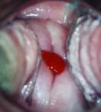
Assessment: may cause inflammatory/impaired-quality screening samples or abnormal bleeding — investigate any abnormal bleeding on its own merits. Differential: a prolapsed endometrial polyp or fibroid (these have a pedicle originating from the uterine cavity, not the cervical canal — refer if in doubt). In postmenopausal women, a cervical polyp warrants USS/hysteroscopy to exclude an associated endometrial polyp.
Associated endometrial polyp occurs in ~27% overall — lower on the COC (~8%), much higher postmenopausally (~57%).
Management: polyps should be removed and sent for histology.
- >1.5 cm → routine gynaecology referral.
- 1–1.5 cm → can be grasped with sponge forceps and twisted off; pressure ± silver nitrate for haemostasis. Removal is usually painless (polyps have no nerve supply); minor bleeding/cramping afterwards. Avoid tampons/intercourse for 2 days; review at 3 months (repeat the screening test if the polyp had affected sample adequacy).
- Recurrence with a normal appearance can be managed the same way (always send histology).
Cervical ectropion (erosion / ectopy)
Glandular (columnar) cells from the endocervical canal present on the outer cervical surface, so the transformation zone is everted and visible. The area looks red (columnar cells are red) at speculum examination.
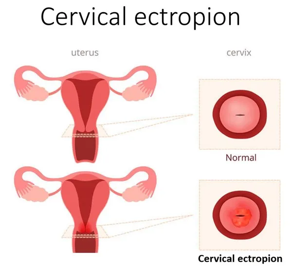
- Cause: hormonal — higher oestrogen states (puberty, pregnancy, the COC). Not linked to cervical cancer or any premalignant condition.
- Usually asymptomatic and self-resolving. Because columnar cells bleed more easily and produce more mucus, it can cause discharge, or bleeding/pain during or after intercourse or a screening test.
- Management: none if asymptomatic. For troublesome symptoms, ablation at colposcopy (silver nitrate or cold coagulation) hardens the columnar cells to stop bleeding. Always exclude other causes of post-coital bleeding first.
Nabothian cyst
Mucinous retention (epithelial inclusion) cysts — a common incidental finding. Form when a cleft of columnar epithelium is covered by squamous metaplasia and the trapped columnar cells keep secreting mucus.
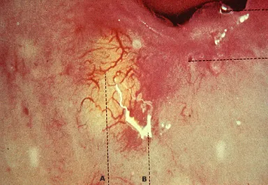
- Discrete cystic structures, microscopic to several centimetres; translucent or opaque; may follow minor trauma or childbirth. Typically asymptomatic.
- Management: none unless symptomatic (pain, a bothersome fullness) or the diagnosis is uncertain. Ablation (electrosurgery) is the usual approach; excise if the diagnosis is unclear to exclude cervical neoplasia. Surgery risks scarring → dyspareunia or cervical stenosis.