Polymyositis / Dermatomyositis
Written 2nd August 2020 - Last Update 23rd July 2026
Polymyositis / Dermatomyositis
Immune-mediated myopathies include dermatomyositis, polymyositis, overlap myositis, necrotising autoimmune myopathy (NAM) and inclusion body myositis (IBM), managed per the British Society for Rheumatology’s dedicated 2022 myositis guideline (registry: bsr-myositis-2022; verified 2026-07-23 — cited by name only, full guideline text not independently fetched).
Classifying immune-mediated myopathies
- Dermatomyositis — adult (with or without malignancy) or juvenile
- Polymyositis
- Overlap myositis — myositis associated with another connective tissue disorder (SLE, mixed connective tissue disease, Sjögren, RA); or antisynthetase syndrome (fever, arthralgia, Raynaud phenomenon, interstitial lung disease, mechanic’s hands — associated with anti-tRNA synthetase antibodies, especially anti-Jo-1)
- Necrotising autoimmune myopathy (NAM) — HMGCR-positive (statin- associated or statin-naive) or SRP-positive; associated with malignancy, connective tissue disease, or viral infection (HIV, hepatitis C)
- Inclusion body myositis (IBM)
- Rarer causes — eosinophilic myositis/myofasciitis, infection-related myositis, granulomatous myositis (including sarcoid-associated)
Clinical features
- Muscle weakness: difficulty raising arms above the shoulders, climbing steps, or rising from sitting; can progress to wheelchair use/bedbound status from pelvic/shoulder girdle weakness. Severe neck flexor involvement can cause inability to lift the head from a pillow. Pharyngeal/upper oesophageal involvement can impair swallowing and cause aspiration risk. Hands, feet and face are spared except in IBM.
- Polyarthralgia or polyarthritis
- Visceral involvement (less common than in other rheumatic disease): interstitial lung disease; cardiac conduction disturbance and ventricular dysfunction; GI involvement (more common in children, via vasculitis — abdominal pain, haematemesis, melaena, ischaemic bowel perforation)
- Skin changes — see dermatomyositis below
Diagnosis
- CK/LDH/AST/ALT elevated
- Electromyography
- MRI — identifies oedema in affected muscle and can help target biopsy
- Muscle biopsy — confirms the diagnosis
Management
Specialist referral — lifelong treatment and monitoring required.
- Exercise under neurophysiotherapy guidance
- First-line: high-dose prednisolone (e.g. 75mg) + a steroid-sparing agent (azathioprine, methotrexate, mycophenolate)
- Severe disease: IV methylprednisolone, IVIg
Dermatomyositis
Muscle inflammation (myositis) with a characteristic cutaneous eruption, which usually precedes muscle weakness onset. Main cutaneous signs:
- Periorbital reddish-purple erythema (heliotrope rash)
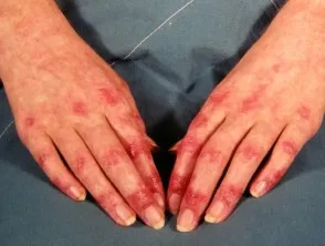
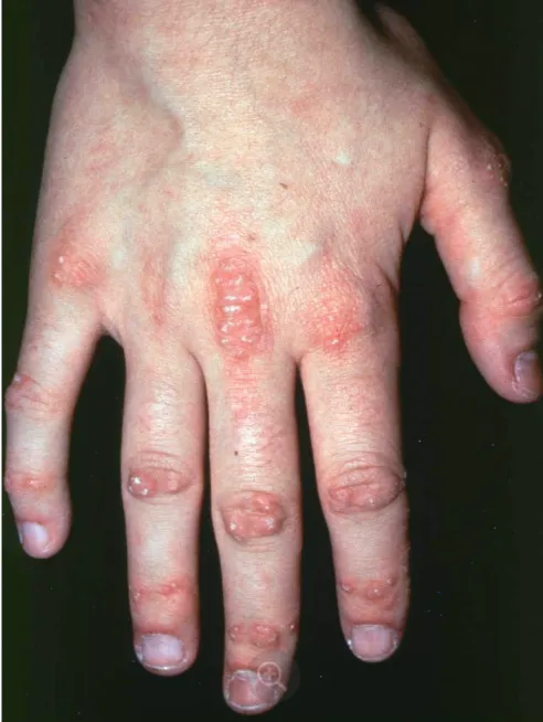
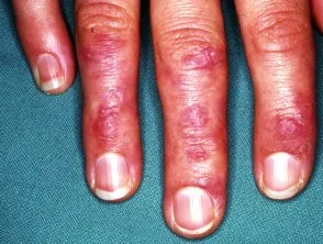
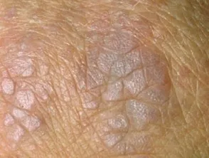
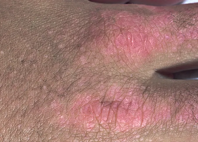
- Flat-topped papules over the dorsal interphalangeal joints (Gottron papules)
- Erythema over the dorsal knuckles (Gottron sign)
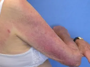
- Photodistributed erythema/oedema on the sun-exposed neck and shoulders (shawl sign)
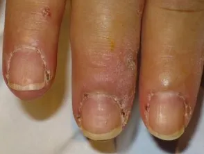
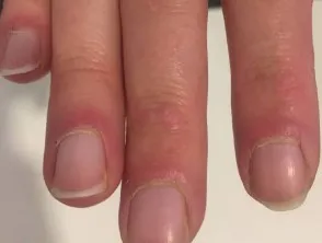
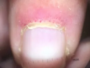
- Characteristic nailfold changes and ragged cuticles
Heliotrope eyelids:
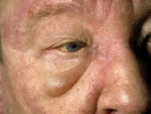
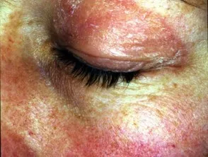
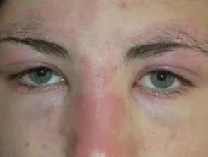
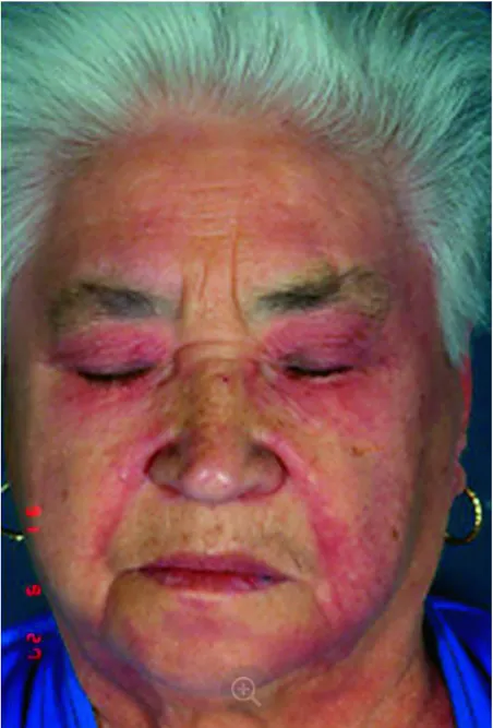
Gottron papules:
Nail folds in dermatomyositis:
Sun-exposed sites:
Dermatomyositis management
- Co-managed by dermatology and rheumatology.
- Topical corticosteroids and hydroxychloroquine are first-line; methotrexate and azathioprine for resistant cases.
- Sun exposure exacerbates disease — stress broad-spectrum sun protection.
- Associated with malignancy in ~25% of adults — consider paraneoplastic dermatomyositis, especially in older patients.
- Dermatomyositis in children is not associated with neoplasia — investigations to exclude occult malignancy are not necessary.