Obstructive sleep apnoea & Sleep disorders
Written 25th January 2020 - Last Update 27th August 2026
Obstructive sleep apnoea & Sleep disorders
Sleep disordered breathing refers to a group of disorders including obstructive sleep apnoea, OSA with a chronic lung disorder, central sleep apnoea, and obesity hypoventilation syndrome.
Obstructive sleep apnoea (adults)
The most common type of sleep disordered breathing, affecting 9–38% of adults. Obesity is the most important risk factor, in a bi-directional relationship/vicious cycle with OSA. Is the presence of apnoeas and hypopnoeas together with daytime sleepiness / dysfunction. Typically patients are overweight, middle-aged, with a thick/short neck and a history of alcohol use and smoking. Recognised risk factors also include being male or a postmenopausal female.
Untreated sleep apnoea is associated with (severe OSA is an independent risk factor): hypertension, arrhythmias (particularly AF), increased cardiovascular mortality, stroke, motor vehicle accidents, and pre-eclampsia/pregnancy-related hypertension. As a memory aid: it’s associated with hypertension, and then whatever hypertension itself causes.
History
Snoring (loud enough to be heard in the next room); waking with choking episodes; morning headaches; witnessed apnoeas; nocturia; nocturnal sweating; sleepiness/poor concentration; GORD; erectile dysfunction.
STOP-BANG
Snoring (louder than talking, or heard in the next room); Tired in the daytime; Observed apnoeas; Pressure (hypertension); BMI >35; Age >50; Neck size (male >43cm, female >41cm); Gender (male).
Low risk = 0–2; intermediate risk = 3–4; high risk = 5+. 4+ needed for medicare funded sleep study (plus ESS >=8).
Epworth Sleepiness Scale (copyrighted…)
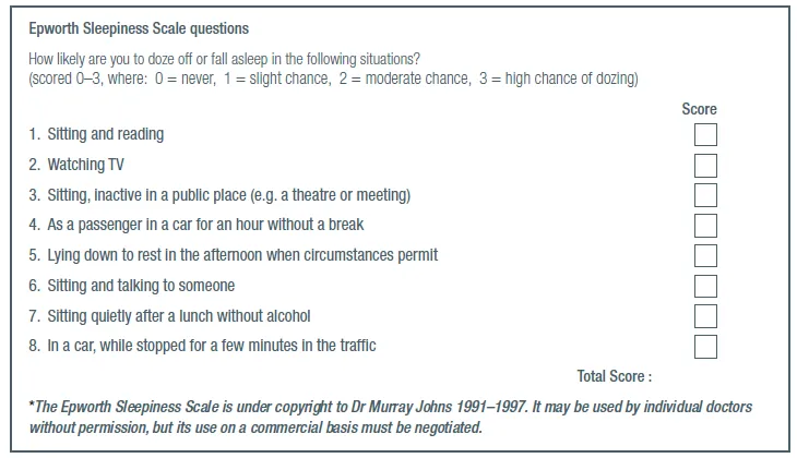
Score 8+ needed for medicare sleep study (+ stopbang 4+)
Requirements for a Medicare-funded sleep study — Epworth Sleepiness Scale score ≥8, plus one of: STOP-BANG score ≥4, OSA-50 score ≥5, or a high-risk Berlin Questionnaire result. A patient is eligible for a Medicare-subsidised diagnostic home-based or laboratory-based sleep study if the Epworth Sleepiness Scale score is 8 or higher plus one of STOP-BANG, OSA-50, or a high-risk Berlin Questionnaire result.
Diagnosis
Standard testing is an overnight sleep study in a lab with polysomnography. General practitioners can also directly request home-based sleep studies, and most patients will be suitable for this option. Home sleep studies have lower sensitivity (but are highly specific). So if home sleep study is negative, then sleep specialist review + laboratory sleep study should be sought if there is high concern about sleep apnoea.
GPs can now order sleep studies directly — a change to Medicare Benefits Schedule rules means a GP no longer needs prior review by a respiratory/ sleep specialist before ordering a diagnostic sleep study, provided the screening criteria above are met. Specialist referral is still appropriate for complex presentations, other suspected sleep disorders, commercial drivers, or patients who fail to comply with/respond to initial treatment.
Patients who meet the Medicare criteria for a sleep study and are referred to the public service will typically have it organised within three months of being referred. Patients with mild to moderate OSA and an Epworth score above 16 who lack severe sleepiness may wait up to three years to be seen in the public sleep service, due to a shortage of specialists.
Comorbid insomnia and sleep apnoea (COMISA) is common and easy to miss — 30–40% of patients with insomnia meet diagnostic criteria for OSA, and 30–50% of OSA patients report clinically significant insomnia symptoms. COMISA causes greater morbidity (daytime impairment, depressive symptoms) than either condition alone and needs both conditions actively managed, not just OSA treatment alone.
Severity grading: Significant sleep apnoeas - Apnoea-hypopnoea index >5/hr; severe >30/hr.
Treatment
General measures
Consider general measures for all patients, but they may be all that is needed in patients with mild disease without daytime symptoms.
- Weight loss (10-15%) — a 10% weight loss produces a clinically meaningful AHI reduction; long-term data suggests roughly 0.78 fewer events/hour for every kilogram lost. GLP-1 receptor agonists are an emerging but not yet established therapy for OSA specifically (as opposed to for weight loss generally) — a tirzepatide trial (SURMOUNT-OSA) was expected to report in 2024 and may change this recommendation; not yet a standard first-line OSA-specific therapy as of this review.
- Avoidance of alcohol and drugs that affect sleep.
- Increased time in bed.
- Positional therapy — to promote side sleeping (pillows not enough); elevate the head of the bed.
- Reduce nasal resistance — smoking cessation, intranasal corticosteroids.
CPAP
The main treatment for OSA is CPAP therapy. CPAP treatment is a significant financial cost and requires ongoing use to effectively manage OSA. CPAP is the gold standard treatment for OSA, improving subjective sleepiness, mood and neurocognitive performance. CPAP also reduces the risk of motor vehicle accidents in patients with OSA. It is the most effective therapy, but needs a titration study organised by a sleep specialist/technicians. Patients who may be eligible for a subsidised CPAP machine should be referred to a sleep disorder specialist to coordinate an application to ENABLE NSW.
Indications: severe OSA; or mild-moderate OSA with daytime dysfunction or hypertension/CVD.
Mandibular advancement splints
Useful in mild-moderate OSA. Needs professional fitment and long term dental review. Can cause TMJ pain; simple mouth guards do not work. Mandibular advancement splints are inferior to CPAP for reducing OSA severity, although better compliance and tolerance may lead to broadly equivalent efficacy in some studies. Mandibular advancement splints are not publicly funded and must be fitted by a dentist with expertise in the area.
Surgery
Rarely required, but may be indicated for significant craniofacial abnormality, tonsillectomy/adenoidectomy if anatomically abnormal, or (very rarely) palatal surgery if anatomically abnormal.
Licensing issues
Pts with OSA have higher rates of motor vehicles accidents. Austroads:
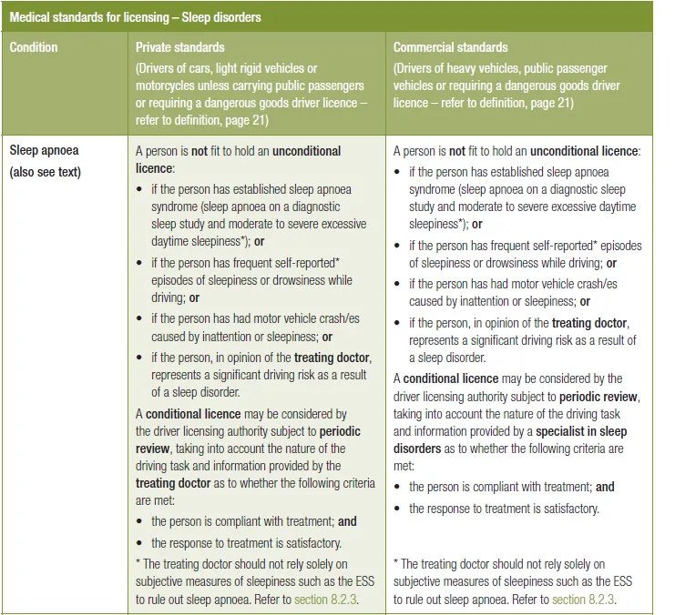
Summary — unfit for an unconditional licence if:
- Established sleep apnoea AND Moderate to severe daytime sleepiness that is untreated.
- Crashes/Frequent incidents secondary to sleepiness whilst driving.
- A patient with frequent self-reported episodes of sleepiness or drowsiness while driving is also not fit to hold an unconditional licence. A patient who has had a motor vehicle crash caused by inattention or sleepiness is similarly not fit to hold an unconditional licence. These patients should not drive until diagnosed, treated, and assessed as suitable for a conditional licence.
Conditional licence OK if: (private licence) the person is compliant with treatment; (commercial licence) compliance AND annual review by a sleep specialist.
See Fitness to Drive (Austroads) for the general private-vs-commercial standards framework and the GP’s reporting duty by jurisdiction.
Obstructive sleep apnoea in children
Commonly due to adenotonsillar hypertrophy. Consider in children who snore or have observed apnoeas when sleeping. These children commonly have hyperactivity, poor school performance and behavioural problems rather than tiredness. Children may also have poor weight gain. Usually occurs between the ages of 2 and 7.
Management: Most children with a suggestive history should just be referred to ENT for consideration for andenotonsillectomy. Sleep studies can be performed if there is doubt.
Central sleep apnoea (CSA)
CSA is a transient loss of the brainstem’s respiratory drive during sleep, rather than an upper-airway obstruction, characterized by transient diminution or cessation of the respiratory rhythm generator located within the pontomedullary region of the brain. Although CSA is less common than OSA, both conditions often coexist, and patients can exhibit features of both states. Symptoms overlap with OSA (poor sleep, morning headache, daytime sleepiness) but snoring is often absent, and patients with CSA are relatively less obese than typical OSA patients. Polysomnography is the diagnostic gold standard.
Causes of brief, periodic loss of respiratory drive: heart failure (Cheyne-Stokes breathing); regular opioid users; premature infants; high altitude.
Causes of prolonged loss of respiratory drive: neuromuscular disease (e.g. MND); chest wall disorder (kyphoscoliosis); severe obesity (OHS).
Several conditions predispose patients to CSA: atrial fibrillation (AF), heart failure (HF) either with preserved or reduced ejection fraction (EF), ischemic stroke, spinal cord injury, renal failure, and chronic opioid use predispose patients to acquire central apnea via transient diminution of ventilatory output — heart failure, opioid use and idiopathic (no identifiable cause) CSA are the commonest patterns seen in practice.
Diagnosis and management require specialist input. CPAP is first-line treatment (as for OSA) and BPAP is a reasonable alternative in hypercapnic CSA unresponsive to CPAP. Adaptive servo-ventilation (ASV) — a form of PAP that responds breath-by-breath to the patient’s own effort — carries a specific, guideline-relevant contraindication: a multicenter largest trial (SERVE-HF) revealed that the use of ASV was associated with increased mortality in HF patients with reduced EF. As a result, ASV is not recommended for the treatment of CSA in this particular group of patients.
Obesity hypoventilation syndrome (OHS)
Sleep-related non-obstructive hypoventilation with CO2 >45mmHg and BMI >35. More precisely, OHS is defined as the presence of awake alveolar hypoventilation characterized by daytime hypercapnia (partial pressure of arterial CO₂ [PaCO₂] >45 mmHg [5.9 kPa]), which occurs as a result of diminished ventilatory drive and capacity related to obesity (BMI >30 kg/m²), without an alternative respiratory, neuromuscular, or metabolic cause for hypoventilation.
Other causes of hypoventilation should be excluded — OHS is a diagnosis of exclusion. Most patients with OHS also have OSA: the majority of patients with OHS (around 70%) have severe OSA, with an AHI of 30 or more events per hour. A minority do not — approximately 10% have OHS without comorbid OSA (also called nonobstructive sleep hypoventilation).
Patients usually have: marked daytime sleepiness; cyanosis/plethora; right heart failure; morbid obesity.
Diagnosis requries sleep study with transcutaneous CO2 measures. A raised serum bicarbonate is a useful screening clue for chronic hypercapnia before that: in patients with serum HCO₃ levels above 27 mEq/L, OHS is present in approximately 50% of cases, while a level below 27 mEq/L makes OHS unlikely if pre-test probability is low.
Treatment is weight loss and CPAP or BiPAP. As with adults’ OSA, PAP therapy, particularly continuous PAP (CPAP), is the first-line treatment for OHS, since most patients have coexisting OSA; BPAP is preferred instead when hypoventilation predominates over obstruction, or when CPAP fails to correct the hypercapnia despite good adherence. Weight loss should not be pursued instead of PAP: treatment should not be delayed while the patient tries to lose weight — a supervised weight-loss target of 25–30% of body weight is recommended alongside PAP therapy, not instead of it. Oxygen alone, without PAP, is strongly discouraged, as it does not improve ventilation and may have poor outcomes with worsening CO₂ retention.
Narcolepsy
Permanent neurological disorder characterised by brief spells of irresistible sleepiness usually at times where an ordinary person feels sleepy. Formally, the only required symptom for the diagnosis of narcolepsy is daily, irrepressible, rapid lapses into REM sleep known as ‘sleep attacks’ that are present for at least three months.
Typically starts between adolescence and 30; close to 50% of patients develop symptoms in their teenage years. It’s rare overall — roughly 14 per 100,000 people have narcolepsy type 1 and around 65 per 100,000 have narcolepsy type 2 disease. Diagnosis is often delayed for years: delays of 5–10 years are common before making a firm diagnosis.
Two types: type 1 (with cataplexy, caused by loss of hypothalamic orexin/hypocretin neurons, thought to be autoimmune) and type 2 (without cataplexy, cause less clear — possibly a milder version of the same orexin loss).
Tetrad of symptoms:
- Daytime hypersomnolence with sleep attacks.
- Cataplexy — sudden, brief muscle weakness triggered by strong emotion (typically laughter). It is pathognomonic for the disorder if it is present but is not required for the diagnosis of narcolepsy type 2.
- Sleep paralysis.
- Hypnagogic hallucinations (vivid, dream-like hallucinations when falling asleep or waking).
Diagnosis is made after excluding chronic sleep deprivation (2 weeks of actigraphy/sleep log), then a polysomnogram followed by a Multiple Sleep Latency Test the next day — a positive test will show a rapid onset of REM sleep (less than 15 minutes) at least twice during the test, and a shortened mean sleep latency averaged across all trials (less than 8 minutes). Type 1 can also be confirmed by a low CSF hypocretin-1 level.
For daytime sleepiness, first-line pharmacologic treatment for excessive daytime sleepiness is modafinil (twice-daily dosing) or armodafinil (once-daily dosing), with amphetamines second-line. For cataplexy, first-line treatment for cataplexy is sodium oxybate (a restricted, centrally-dispensed medication), with tricyclic antidepressants or SNRI/SSRIs also used with some success for cataplexy, sleep paralysis and hypnagogic hallucinations. Scheduled short naps (15–20 minutes) and good sleep hygiene are useful adjuncts. In children, currently, there is no medication that has been approved by the FDA for use in children.
Restless leg syndrome
Prevalence increases with age, mainly affecting elderly people. Diagnosis is made from history — there are no special diagnostic tests. Secondary causes include anaemia (common), iron deficiency (common), uraemia, hypothyroidism, pregnancy (usually ceases within weeks of delivery), and drugs (antihistamines, anti-emetics, lithium, antidepressants, antihypertensives and tranquillisers).
Management — non-pharmacological
- Self-help advice.
- Perform activities that can reduce symptoms (e.g. Modest amount of walking before bedtime, massage or stretching exercises for hamstring and calf muscles for 5 mins before bed).
- Good sleep hygiene.
- Diet: avoid caffeine, smoking and alcohol.
- Try keeping legs cooler than the body for sleeping.
Management — pharmacological
The 2024 AASM guideline reverses the medication ladder below: check serum ferritin and transferrin saturation, replacing iron if ferritin is low before starting any of these drugs, and prefer an alpha-2-delta ligand (gabapentin, gabapentin enacarbil, or pregabalin) first-line — the AASM recommends the use of gabapentin enacarbil over no gabapentin enacarbil as a strong recommendation. Dopaminergic agents are no longer preferred long-term therapy (see below).
- Clonazepam 0.5–1mg, 1 hour before bed.
- Levodopa (+ benserazide or carbidopa) 100–200mg — the AASM suggests against the standard use of levodopa due to augmentation risk; may still suit patients wanting short-term symptom control who accept that risk.
For more severe symptoms, consider low dose dopamine agonists:
- Pramipexole 0.125mg daily, up to 0.75mg — the AASM suggests against the standard use of pramipexole for the same augmentation-risk reason (ropinirole and rotigotine carry the same caution).