Renal Cancer / Incidental Renal Mass
Written 26th August 2020 - Last Update 19th August 2026
Renal Cancer / Incidental Renal Mass
Renal cell carcinoma (RCC) represents 2–3% of all cancers, and more than 50% are detected incidentally (on imaging done for another reason).
Epidemiology
- Lifetime risk 1 in 74; male:female ~2:1; peak incidence 60–70 years
- Established risk factors include smoking (dose-dependent, roughly doubles risk), obesity and hypertension; other associations include occupational/environmental exposures (asbestos, cadmium, petroleum products) and familial syndromes (e.g. von Hippel–Lindau, tuberous sclerosis)
- Inherited forms account for ~3% of RCC — ask about family history
Small renal masses
Defined as <4cm (stage T1a by definition). ~20% are benign; 20–25% prove to be potentially aggressive at diagnosis.
Clinical presentation
Examination is usually unremarkable — the classic triad of flank pain + gross haematuria + abdominal mass is uncommonly seen. Ask about and examine for paraneoplastic features: hypertension, anaemia, hypercalcaemia, cachexia, pyrexia, weight loss, polycythaemia, raised ESR, abnormal liver enzymes.
Investigations
- UEC + eGFR, FBC, ESR, ALP, LDH, corrected serum calcium
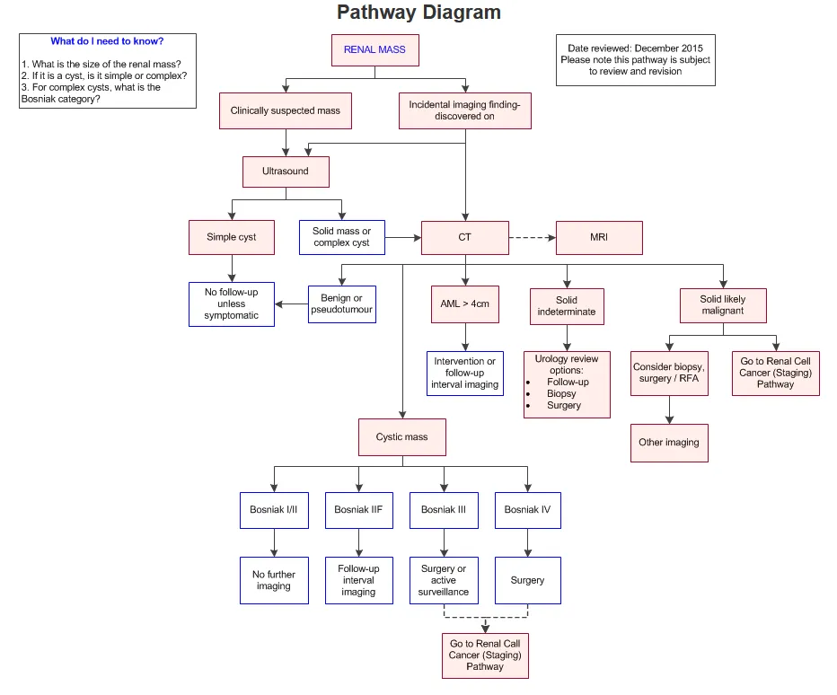
- Four-phase contrast CT is the best imaging modality
- For cystic masses, the Bosniak classification (see
renal-cysts.md) predicts malignancy risk from CT appearance — thickened/irregular septa and enhancement are concerning features
The role of biopsy
~20% of lesions highly suspicious for RCC on imaging prove benign after surgery. CT-guided biopsy is safe — minimal bleeding risk, and needle-tract tumour seeding is extremely rare. The decision to biopsy should be made by a urologist.
Management
Surveillance vs surgery vs ablative techniques is a urologist-led decision. Surgical resection is the treatment of choice for localised RCC, but surveillance of a small RCC may be preferable in older patients with significant comorbidity given surgical risk.