Strabismus
Written 17th April 2020 - Last Update 1st August 2026
Strabismus
Common disorder of ocular alignment affecting 2–4% of children, commonly known as squint or lazy eye. There is no national Australian screening programme for strabismus, so early detection depends on opportunistic pick-up by GPs and maternal/child health nurses.
The most severe consequence is amblyopia — permanent loss of best-corrected visual acuity in a structurally healthy eye, caused by abnormal visual experience during critical periods of early neurodevelopment downregulating the neural pathways to the visual cortex. After around age 9 these pathways may not recover even if normal visual function is later restored. Strabismus also carries a lifelong cosmetic and psychosocial burden — poor self-esteem, social prejudice and restricted career opportunities have all been described. Early detection and treatment improves both visual and psychosocial outcomes.
Differential diagnosis: is this actually strabismus?
Two common look-alikes should be considered before referring:
- Ocular instability of infancy — newborns often have unsteady ocular alignment for up to 3 months of age, usually a lateral deviation of less than 15°. This is a normal finding and does not need referral.
- Pseudostrabismus — unusual facial architecture (e.g. a broad nasal bridge/telecanthus, or prominent epicanthal folds obscuring the nasal sclera) creates the visual illusion of an eye turn on light-reflex/red-reflex testing when the cover test is actually normal. If in doubt, refer for specialist assessment.
Any ocular deviation greater than 15°, or one persisting beyond 3 months of age, is considered abnormal and warrants assessment.
Common causes of strabismus
| Primary causes of strabismus | Secondary causes of strabismus |
|---|---|
Idiopathic strabismus (risk factors: family history, prematurity, low birth weight) Congenital syndromes |
Cranial nerve palsies (CNIII, IV, VI) Orbital fracture Intracranial bleed Intracranial/intraorbital/intraocular mass (benign or malignant) Intracranial infection Grave's disease Myasthenia gravis Diabetes mellitus Amblyopia and uncorrected refractive error (a degraded image in one eye can itself cause the eye to drift) Toxins and heavy metal poisoning Post-vaccination |
Types of strabismus
Most common is horizontal misalignment, but vertical misalignment also occurs. Ocular deviation may be manifest (tropia — constant, intermittent, or fluctuating and worse with fatigue) or latent (phoria). It can present in all directions of gaze (comitant) or only in a specific direction (incomitant).
Symptoms can vary over time — deviation that seems well controlled in early childhood may become more apparent as the child grows, so ongoing evaluation is needed even after an initial reassuring assessment.
| Terminology |
|---|
eso- = Nasal horizontal deviation (relative to fixing eye) exo- = Temporal horizontal deviation (relative to fixing eye) hyper- = Superior vertical deviation (relative to fixing eye) hypo- = Inferior vertical deviation (relative to fixing eye) tropia = Manifest disorder of ocular alignment phoria = Latent disorder of ocular alignment comitant = Ocular deviation present in all directions of gaze incomitant = Ocular deviation only present in specific directions of gaze amblyopia = Clinically defined as a 2-line difference from best corrected visual acuity in a structurally healthy eye |
History
Aims to distinguish primary from secondary causes and to screen for red flags suggesting recent trauma or serious intracranial pathology. New strabismus in a school-aged child is uncommon and warrants further investigation.
Take a focused obstetric, developmental and medical history — malignancy, autoimmune conditions, exposures, vaccinations, and the child’s general health/any unexplained illness. Ask about onset and any associated signs such as gait disturbance, nystagmus, reduced visual acuity or diplopia, and any history of head/orbital trauma.
Screening tests
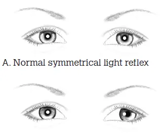
Light reflex test — from 1 m with a small light, compare the light’s reflection in both eyes. Identical reflection is normal; deflection indicates abnormal alignment (each 1 mm of deflection ≈ 15–20 prism dioptres of deviation).
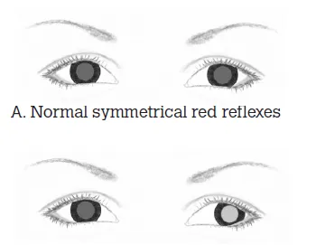
Red reflex test — at 0.5 m with a direct ophthalmoscope set to zero power, view the red reflex in both eyes simultaneously. Should be identical in size, shape and colour.
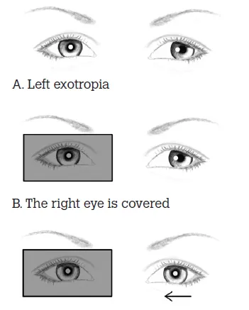
Cover test — direct the child’s attention to a target, then cover one eye while observing the uncovered eye. Movement of the uncovered eye to take up fixation indicates manifest strabismus (tropia) in that eye. Repeat on each eye. This test also distinguishes true strabismus from pseudostrabismus.
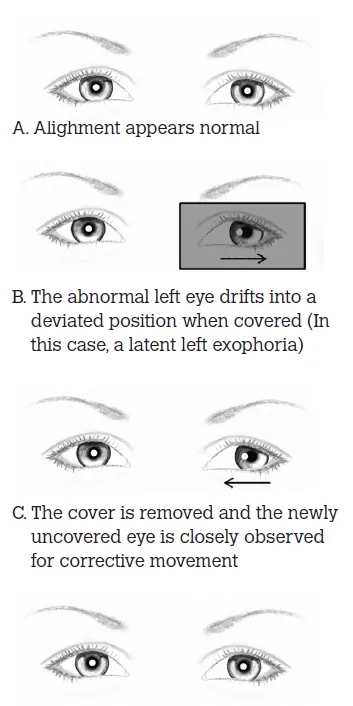
Uncover test — cover one eye for 5 seconds, then remove the cover and observe the newly uncovered eye. A latent deviation drifts under cover then corrects back on uncovering, indicating a phoria in that eye.
Management and referral
Any red flag on history or examination (acute onset, trauma, neurological signs, school-age new onset) needs urgent same-day specialist discussion. Otherwise, urgency depends on the type and pattern of deviation. RCH’s pre-referral pathway (still the standard reference triage framework) gives concrete thresholds:
| Presentation | Action |
|---|---|
| Intermittent/variable esotropia before 3 months of age | Normal for infants — do not refer |
| Intermittent/variable esotropia after 3 months of age, or constant esotropia at any age | Refer semi-urgently for evaluation |
| Constant esotropia after age 9, longstanding | Refer routinely to local optometrist/ophthalmologist |
| True acute-onset constant esotropia, any age | Immediate referral — same-day ophthalmology contact |
| Intermittent or constant exotropia up to age 9 | Refer routinely |
| Exotropia after age 9 | Refer to local optometrist/ophthalmologist |
| True acute-onset constant exotropia | Immediate referral — same-day ophthalmology contact |
All other newly diagnosed strabismus still warrants timely (non-immediate) ophthalmology referral for comprehensive assessment. Treatment depends on primary vs secondary cause and may include refractive correction (glasses/contacts), amblyopia therapy (patching or atropine penalisation), prisms, and surgical correction (common, well tolerated, day-case). Goals are to prevent/treat amblyopia and achieve binocular vision with functional depth perception, with better cosmesis a secondary goal.