Congenital Heart Disease
Written 8th April 2020 - Last Update 28th August 2026
Congenital Heart Disease
See also murmurs in childhood for the innocent-murmur screen and cardiac history/exam red flags that apply across every lesion below.
Acyanotic lesions (left-to-right shunts)
Atrial septal defect
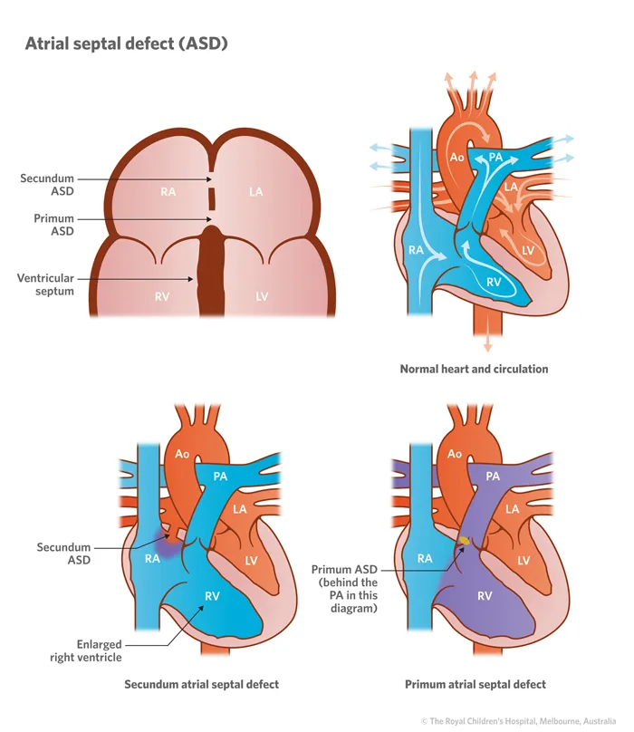
An abnormal connection between the two atria. Two types:
- Ostium secundum — higher in the septum, less severe.
- Ostium primum — lower in the septum, more severe.
History: ostium secundum: symptoms are uncommon in infancy; ostium primum: heart failure with pulmonary hypertension develops early.
Examination: mid-systolic murmur in the pulmonary area; loud P2 with widened splitting of its pulmonary component.
Investigations: echocardiogram.
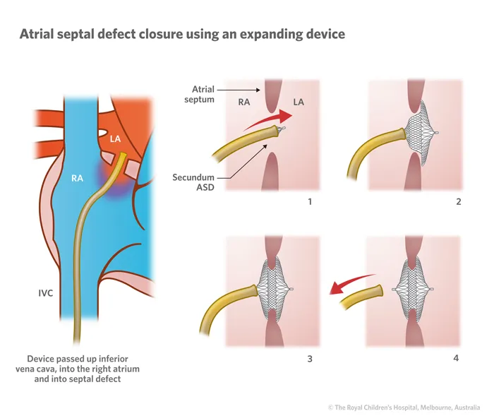
Management: refer to cardiology for advice — closure may be considered if there’s troublesome shunting. Some are monitored with serial echocardiograms for spontaneous closure. Delayed growth/development makes closure more advisable. Endocarditis prophylaxis is not needed preoperatively and is required only for the first 6 months after repair or if there is a residual defect adjacent to a surgical patch.
Ventricular septal defect
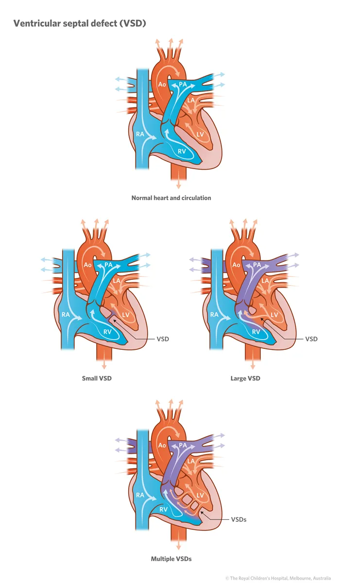
Connection between the two ventricles with a left-to-right shunt. The most common congenital heart lesion (~1 in 500 births).
History: large VSD — symptoms appear in infancy: breathlessness on feeding or crying, recurrent chest infections, failure to thrive; heart failure develops from around 3 months.
Examination: palpable thrill at the left sternal edge; pansystolic murmur at the right sternal edge. A smaller VSD produces a harsher murmur (higher-velocity jet through a tighter defect).
Investigations: echocardiogram.
Management: refer to cardiology early** for consideration of primary closure — but ~50% close spontaneously.
Patent ductus arteriosus
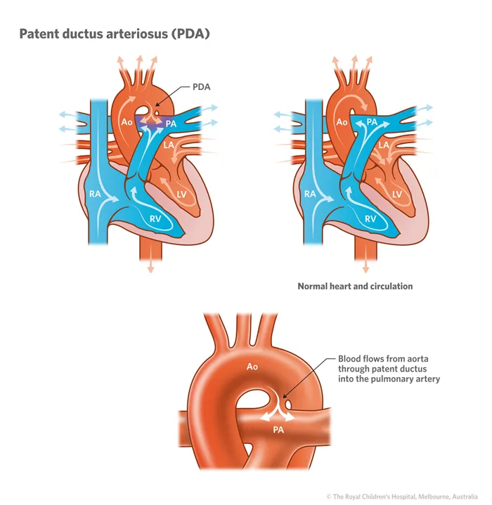
The ductus arteriosus normally closes in the first few weeks of life. Failure of closure leaves a shunt from the aorta to the pulmonary artery.
Examination: loud, continuous “machinery” murmur. Symptoms and signs relate to shunt size — a large shunt presents with failure to thrive, respiratory infections and heart failure.
Investigations: echocardiogram.
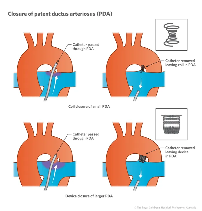
Management: refer to cardiology — closure is considered if symptomatic. Monitor for signs of infection, as these children are at increased endocarditis risk.
Coarctation of the aorta
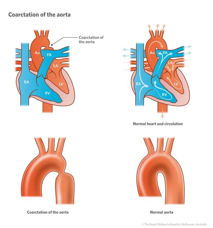
Congenital narrowing of the aorta.
History: usually presents in infancy with heart failure as the ductus arteriosus begins closing (day 4–10) — blood pressure elevated in the arms, reduced in the legs, with reduced femoral pulses. Presenting later in childhood, the child is hypertensive with reduced leg pulses. Four-limb blood pressure and femoral/brachial pulse comparison are the key screening manoeuvres (see murmurs in childhood).
Management: most cases need surgical repair — refer.
Cyanotic lesions (right-to-left shunts)
The major right-to-left shunting congenital lesions (“the 5 T’s” plus tricuspid/pulmonary atresia).
Tetralogy of Fallot
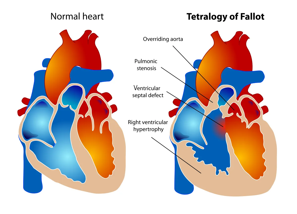
The key components are pulmonary artery stenosis and a VSD, which together create the right-to-left shunt and cyanosis; the aorta overrides the VSD (sits astride it).
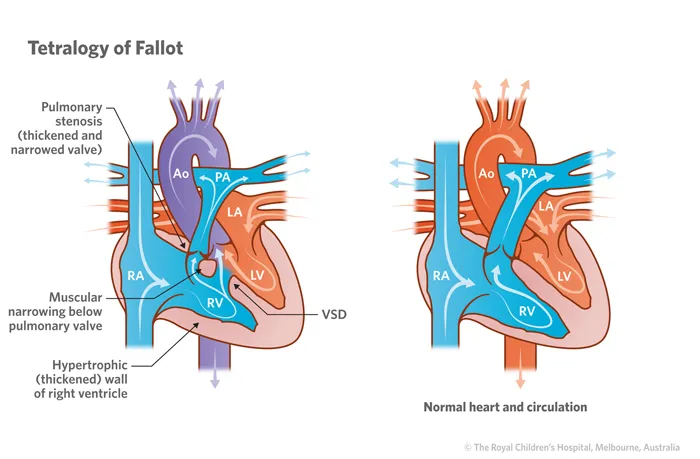
- History: cyanosis is not typically seen in the newborn period — appears at 6–12 months, more apparent with crying/exertion; may present later if shunting is less severe. “Tet spells” — sudden pallor and cyanosis without obvious cause, the child appearing unwell or floppy then recovering spontaneously. Progressively reduced exercise capacity.
- Examination: harsh ejection systolic murmur at the left sternal edge; loud second heart sound; clubbing develops over time; sometimes growth restriction.
- Investigations: CXR may show a “boot-shaped” heart; ECG shows RV hypertrophy; echocardiogram is diagnostic.
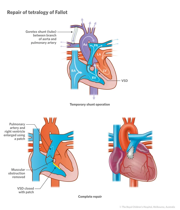
- Management: surgical repair of the VSD and pulmonary stenosis. Beta-blockers (e.g. propranolol) can reduce hypoxic spells while awaiting surgery. Many repaired patients eventually need pulmonic valve replacement.
Transposition of the great arteries
The pulmonary artery and aorta are swapped, creating two separate (parallel) circuits. Survival depends on mixing between the circuits via a PFO, the ductus arteriosus, or a septal defect.
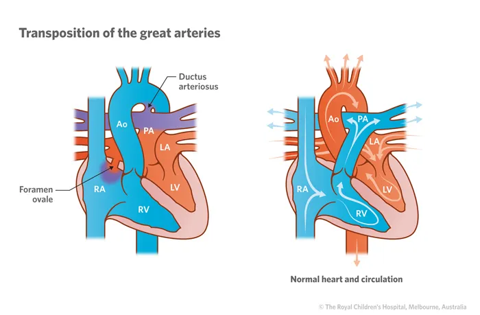
- History: cyanosis presents in the first few hours of life, progressing over subsequent days.
- Examination: possible RV heave at the left sternal edge; usually no murmur.
- Investigations: echocardiogram is essential.
- Management: initial balloon atrial septostomy creates an ASD to buy time, then definitive surgery — the arterial switch operation (arteries and coronary arteries moved to the correct side) — usually performed in the first few weeks of life.
Tricuspid atresia
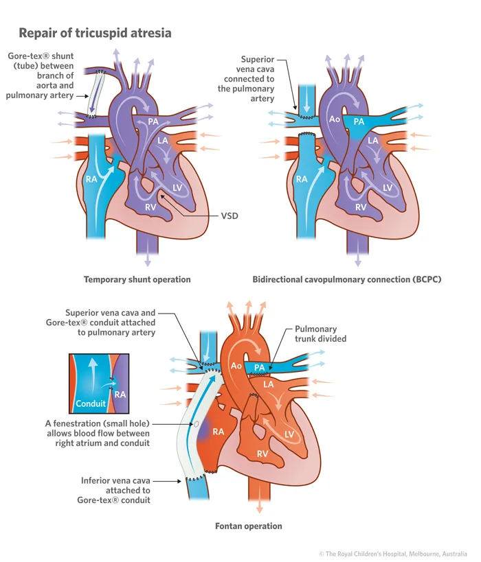
Complete tricuspid valve blockage — blood passes RA→LA via the foramen ovale or a septal defect, then reaches the pulmonary circulation via the ductus arteriosus, giving very limited pulmonary flow.
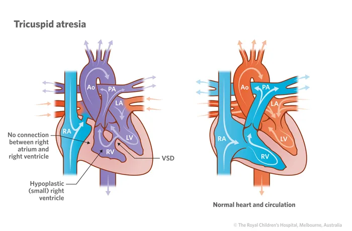
- History: early cyanosis. Examination: systolic murmur.
- Investigations: echocardiogram (also characteristic ECG/CXR findings).
- Management: complex staged surgery — initially a shunt connecting the pulmonary artery to the subclavian artery, then definitive correction by connecting the SVC/IVC directly to the pulmonary arteries (Fontan operation).
Pulmonary atresia
Complete blockage or absence of the pulmonary artery origin — blood flows right-to-left via the foramen ovale or a septal defect.
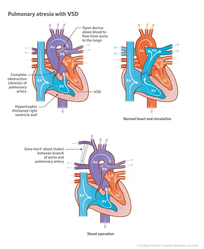
- History: early cyanosis. Examination: continuous murmur from a PDA.
- Investigations: echocardiogram (characteristic complex ECG/X-ray findings).
- Management: prostaglandin infusion to keep the ductus arteriosus open, then a systemic-to-pulmonary shunt, with later correction by inserting a pulmonary valve conduit.
Truncus arteriosus
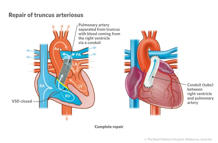
A single arterial trunk arises from both ventricles and then splits into the aorta and pulmonary arteries.
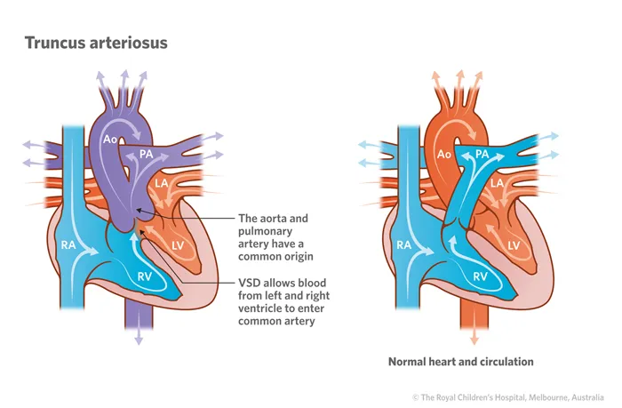
- Examination: cyanosis is mild or even absent; congestive heart failure occurs early in infancy (hepatomegaly, breathlessness on feeding/crying); systolic ± diastolic murmur (from truncal valve regurgitation).
- Investigations: echocardiogram.
- Management: surgery to separate the arterial trunks and place a valved conduit to the pulmonary circulation.