Uveitis
Written 15th July 2020 - Last Update 23rd July 2026
Uveitis
Inflammation of the uveal tract — the vascular coat of the eye, comprising the iris, ciliary body and choroid. Uveitis may be anterior or posterior.
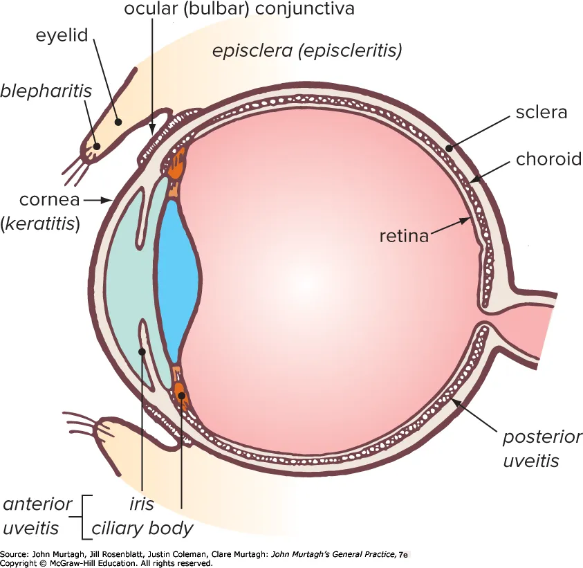
Anterior uveitis (acute iritis)
Inflammation of the iris and ciliary body — the iris becomes sticky, adhering to the lens and losing its light response.
Kumar & Clark triad: redness, pain, photophobia.
Causes:
| Category | Examples |
|---|---|
| Autoimmune | Seronegative arthropathies, SLE, inflammatory bowel disease, sarcoidosis |
| Infectious | Toxoplasmosis, syphilis, herpes viruses (including CMV), HIV |
History: red eye (especially around the iris), eye pain, photophobia, blurred vision, floaters, small pupil, tearing.
Examination: constricted, irregular pupil; hypopyon (pus in the anterior chamber) may be present.
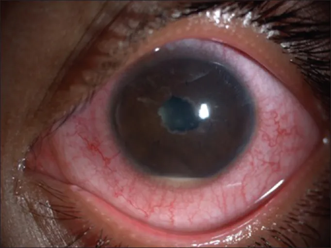
Investigations: slit lamp examination + tonometry (to differentiate from glaucoma).
Management:
- Seen in ED for slit lamp exam + tonometry to confirm diagnosis; ophthalmology review within 24 hours
- Pupil dilation (e.g. atropine eye drops) to prevent/break synechiae
- Topical steroids to reduce inflammation ± systemic steroids
Prognosis: good if treated early and consistently, but recurrence is likely.
Posterior uveitis (choroiditis)
Inflammation of the choroid, which may involve the retina and vitreous.
History: no pain, blurred vision, floaters (floating opacities).
Management: referral to ophthalmology.