Scleritis / Episcleritis
Written 16th July 2020 - Last Update 23rd July 2026
Scleritis / Episcleritis
Both cause a red eye without discharge, but scleritis is a severe, vision-threatening emergency while episcleritis is benign and self-limiting. Distinguishing them matters.
Scleritis
Severe, painful, vision-threatening inflammation of the sclera and deep episclera.
Associated systemic conditions: rheumatoid arthritis, herpes zoster, sarcoidosis, tuberculosis — and other systemic vasculitides.
History: red eye that is more violaceous (blue-red/purple) than episcleritis or conjunctivitis; deep, boring pain severe enough to interfere with sleep; ± photophobia and lacrimation.
Examination:
- Redness may be segmental or involve the whole globe; palpebral conjunctiva is normal
- Lacrimation without discharge (distinguishes it from conjunctivitis)
- A raised nodule (nodular scleritis) or area of necrosis (necrotising scleritis) may be seen
- The globe is tender to palpation
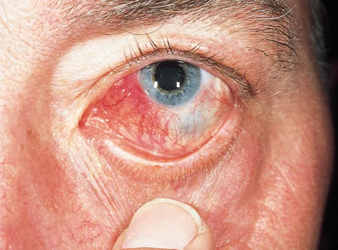
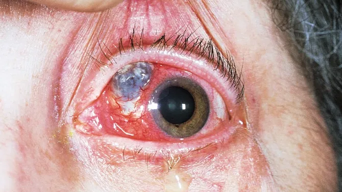
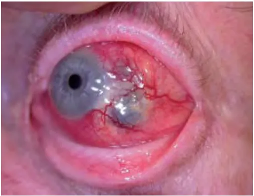
Investigations: clinical diagnosis, but requires slit lamp examination.
Management:
- Urgent referral — ophthalmology review within 24 hours
- NSAIDs for mild cases (rarely sufficient alone)
- Systemic steroids: prednisolone 1–2 mg/kg for 7 days then taper
- Immunosuppressants for unresponsive cases
- Refer for investigation of an underlying rheumatological condition — commonly present
Prognosis: significant risk to vision. Necrotising scleritis can progress to globe perforation, needing surgical repair or enucleation. 14% lose significant visual acuity at 1 year, 30% at 3 years. Necrotising scleritis carries a 50% 10-year mortality, largely from myocardial infarction related to the underlying systemic vasculitis.
Episcleritis
Recurring, self-limiting inflammation of the episclera (the thin vascular membrane between conjunctiva and sclera) that does not threaten vision.
Demographics: young adults, women > men.
History: localised redness, lacrimation, usually mild irritation. Distinguished from scleritis by the absence of photophobia/severe pain, and from conjunctivitis by the absence of discharge.
Examination: segmental hyperaemia, less violaceous than scleritis; no palpebral conjunctival involvement.
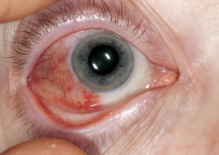
Investigations: clinical diagnosis with slit lamp examination.
Management: topical corticosteroid eye drops, ideally in consultation with ophthalmology.