Retinitis Pigmentosa
Written 18th July 2020 - Last Update 15th August 2026
Retinitis Pigmentosa
A group of inherited degenerative retinal diseases causing night blindness and progressive vision loss. Also called RP, pigmentary maculopathy, pigmentary retinopathy, or rod-cone dystrophy. Caused by abnormal melanin pigmentation in the retina, some of which is also found floating in the vitreous.
Types
- Non-syndromic (“simple”) — doesn’t affect other organs/tissues; multiple inheritance patterns and genetic mutations
- Syndromic — a component of ~50 recognised syndromes; 20–30% of RP patients have other systemic symptoms
- Systemic — affects multiple tissues/organs
Epidemiology
- Prevalence: 1 in 3,000–7,000 people (14–33 per 100,000); ~1 in 4,000 in the US, ~1 in 5,000 worldwide
- Mean age at diagnosis 35 years
- A leading cause of blindness (29%) in patients under 60–65 in some cohorts (Kuwait, Denmark)
- 15–25% of cases are autosomal dominant — genetic screening should be offered to all relatives given this
Genetics
- 40–50% of cases have no identified genetic cause
- Autosomal dominant (15–25% of cases) — rhodopsin (RHO) is the most common gene (25–30% of AD cases)
- Over 80 genes implicated, causing defects in the phototransduction cascade, vitamin A metabolism, protein transport, or other cellular functions — leading to progressive photoreceptor death
Associated syndromes (recognise the pattern, detail not essential)
Usher syndrome (types I–III, RP + hearing loss ± vestibular dysfunction — the most common syndromic association, ~18% of all RP), Alström syndrome, Refsum disease (RP + anosmia + neuropathy + deafness + ataxia + ichthyosis — treatable with dietary phytanic acid restriction), abetalipoproteinaemia/Bassen-Kornzweig (treatable with vitamin A/E), Bardet-Biedl syndrome, gyrate atrophy, choroideremia, cone-rod dystrophy, Leber congenital amaurosis, Senior-Løken syndrome (RP + renal failure), mitochondrial disorders.
Presentation
- Night blindness (nyctalopia) and impaired dark adaptation — usually begins in adolescence
- Followed by mid-peripheral visual field loss in young adulthood — often unnoticed by the patient until it progresses (“tunnel vision”); patients may just seem “clumsy” from the restricted field
- Central visual acuity is usually preserved until late in the disease course
- Ask about visual problems in all first-degree relatives across 3 generations
Examination
- Usually bilateral (unilateral RP is possible)
- Early: fundus may appear normal (“RP sine pigmento”)
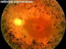
- Progressive fundus changes: arteriolar narrowing → fine “dust-like” intraretinal pigmentation → coarse melanin clumping (“bone spicule” appearance) → attenuated retinal vessels, pale/waxy optic nerve
- Posterior subcapsular cataracts in up to ~70%, severity correlating with age
- Visual field: a ring scotoma ~20–25° from fixation in early disease, progressing outward slowly and inward faster → tunnel vision; may meet legal-blindness criteria (peripheral field ≤20° diameter/≤10° radius bilaterally) despite preserved acuity
Diagnosis
1983 consensus criteria: rod dysfunction (elevated dark-adaptation threshold or abnormal electroretinogram), progressive night blindness, progressive peripheral field loss, and bilateral involvement.
Management
Goal is slowing decline, not cure, for the great majority of cases:
- UV-blocking sunglasses (protective)
- Higher dietary omega-3 intake — associated with slower visual acuity decline
- Vitamin A palmitate 15,000 units/day — may slow decline in cone ERG amplitude
- Lutein supplementation — may slow visual field decline
- Avoid high-dose vitamin E (400 IU/day) — may increase visual decline (cone ERG amplitude)
- Acetazolamide for acute cystoid macular oedema episodes only (not for chronic disease)
- Cataract surgery if visually significant
- Laser/cryotherapy if inflammation leads to retinal detachment
- Argus II Retinal Prosthesis System — FDA-approved implantable device for advanced RP
- Genetic counselling and psychological support (major disease milestones, chronic disability adjustment)
Prognosis
- Most patients become legally blind at some point in the disease course; however, total (light-perception) vision loss is uncommon — many retain good central vision into their 40s–50s, since macular function is often preserved even after peripheral field is lost
- Course is highly variable and genotype-dependent — X-linked disease in males tends to be worst; autosomal dominant disease tends to be best; Bassen-Kornzweig, Refsum disease and isolated vitamin E deficiency have comparatively better outcomes (and are the treatable subtypes above)
Pregnancy
5–10% of pregnant women with RP report decreased vision that frequently does not recover postpartum — possibly related to pregnancy-associated corneal/lens changes.
Screening
Genetic testing for relatives of affected individuals; antenatal diagnosis (amniocentesis/ chorionic villus sampling) can identify an affected fetus. Note that clinical severity and phenotype can differ even with the same genetic variant, so a relative’s disease course doesn’t reliably predict another’s.
Sources: https://www.healthdirect.gov.au/retinitis-pigmentosa, https://www.betterhealth.vic.gov.au/health/conditionsandtreatments/eyes-retinitis-pigmentosa