Retinal Vascular Occlusions: Artery & Vein
Written 20th July 2020 - Last Update 28th August 2026
Retinal Vascular Occlusions: Artery & Vein
The two central retinal vascular occlusion syndromes — arterial (an acute ischaemic-stroke equivalent) and venous (a more indolent, often-improving process) — covered together for comparison.
Central retinal artery occlusion (CRAO)
Presents with sudden, profound loss of vision.
Causes:
- Most frequently emboli obstructing the retinal artery (e.g. cardiac/carotid source, as in stroke)
- Less commonly, in-situ thrombus formation from vasculitis (giant cell arteritis, SLE)
Examination:
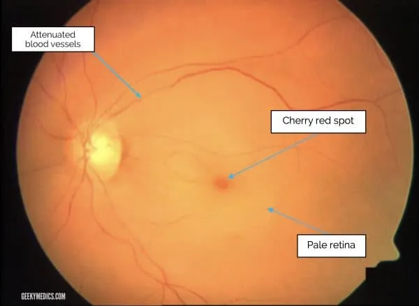
- Cherry-red spot on the fovea — the intact foveal reflex standing out against the pale, ischaemic retina
- Relative afferent pupillary defect (RAPD)
Investigations:
- ESR/CRP/platelets — screen for giant cell arteritis (see
abnormal-eye-movements-diplopia.md’s GCA section) - Once diagnosed, look for an embolic source: carotid duplex USS (carotid artery stenosis), echocardiogram (transmural thrombus, atrial myxoma)
Treatment: CRAO is now managed as an acute ischaemic stroke equivalent. A 2021 AHA scientific statement reframes non-arteritic CRAO this way — IV thrombolysis (alteplase) within a 4.5-hour window may improve visual recovery and is now used at stroke-capable centres, though the evidence base remains limited. This means CRAO should trigger urgent stroke-pathway activation / ED transfer rather than being treated as a low-yield, ophthalmology-only presentation — early treatment is key to any chance of vision preservation.
- If GCA is the underlying cause, steroid treatment can prevent vision loss in the fellow eye, and may recover some vision in the affected eye too
- Older adjunct measures (IOP reduction with timolol/acetazolamide) are of limited proven benefit beyond the acute stroke-pathway window but may still be trialled. Mechanical embolectomy is not feasible given the small calibre of the retinal/ophthalmic vessels — intra-arterial thrombolysis remains unproven but may be attempted at centres capable of endovascular therapy.
Central retinal vein occlusion (CRVO)
Painless vision loss, usually sudden but may progress over days to weeks.
Risk factors: hypertension (major), age (major), glaucoma, diabetes, increased blood viscosity.
Mechanism: occurs from atherosclerotic thickening of the central retinal artery compressing the central retinal vein at their shared crossing point. Venous occlusion causes retinal hypoxia; the resultant endothelial cell damage leads to blood extravasation.
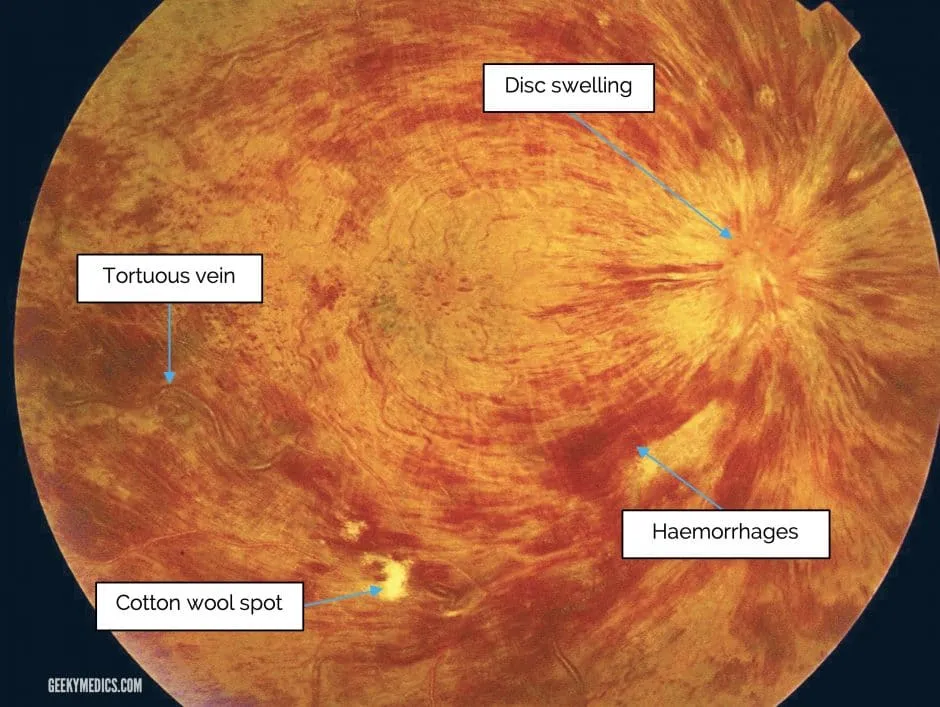
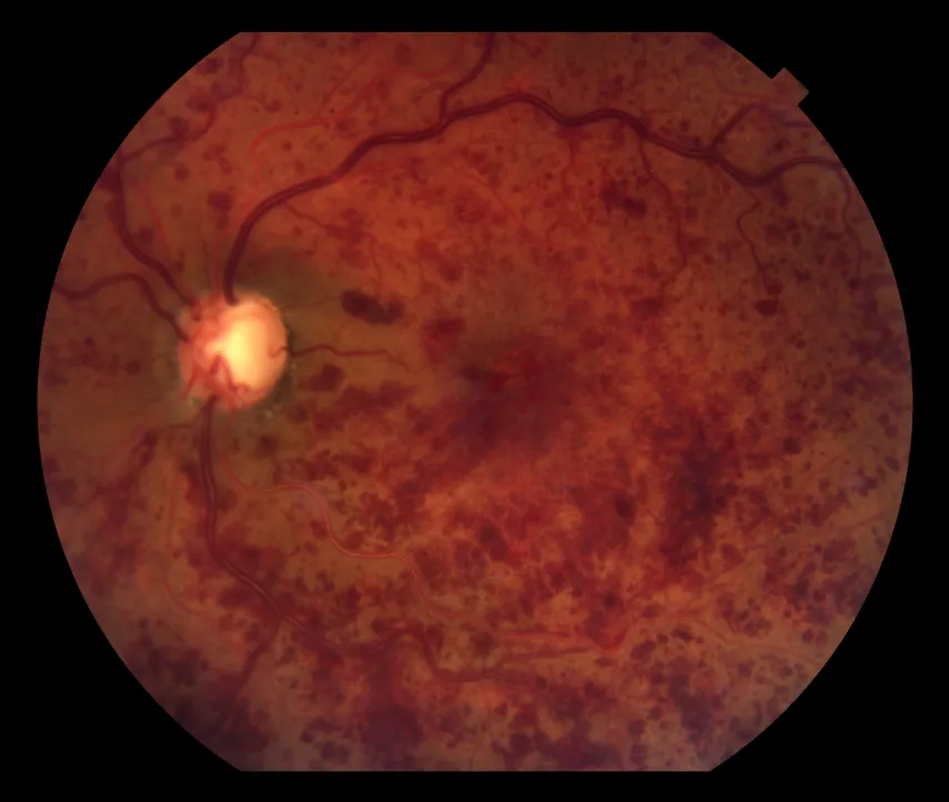
Examination: fundoscopy typically shows severe tortuosity and engorgement of retinal veins, deep haemorrhages, cotton wool spots, and optic disc swelling.
Prognosis: correlates with presenting vision — spontaneous improvement is more likely the less severe the initial deficit:
- Vision ≥6/12 at presentation → improvement likely
- Vision 6/60 → improvement unlikely, may potentially worsen
Treatment:
- Intravitreal injections — anti-VEGF or dexamethasone
- Laser photocoagulation