Retinal Detachment
Written 16th July 2020 - Last Update 28th August 2026
Retinal Detachment
A vision-threatening emergency requiring timely recognition and referral. Affects approximately 1 in 10,000 Australians annually. Painless vision loss from physical separation of the neurosensory retina from the underlying retinal pigment epithelium, requiring surgical repair.
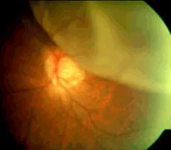
Rhegmatogenous retinal detachment (the most common type) occurs when fluid from a vitreous/retinal tear passes underneath the retina, separating it from the retinal pigment epithelium and causing progressive detachment. If the macula is involved, central vision loss is usually permanent. Patients may describe a curtain coming down over their vision as the peripheral fields detach.
Retinal tear
Usually caused by posterior vitreous detachment — vitreoretinal traction during PVD can tear
the retina, allowing subretinal fluid to accumulate and progress to retinal detachment (see
posterior-vitreous-detachment.md for the fuller PVD picture).
Presentation:
- No visual field defect or relative afferent pupillary defect
- Black or red spots may be seen if there has been bleeding
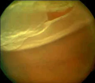
Treatment: Prompt treatment is needed to prevent progression to retinal detachment:
- Laser or cryotherapy burns create a chorioretinal scar that seals the tear and prevents fluid tracking underneath the retina
The “4 Fs” — hallmark warning symptoms
Flashes, Floaters, Field defect, Failing vision. None is pathognomonic alone, but any combination — or any of them in a higher-risk patient — warrants urgent ophthalmic evaluation. A field defect is almost pathognomonic for retinal detachment. Failing vision (any acute vision loss) is a red flag in any presentation.
Quantified risk factors
| Risk factor | Relative risk |
|---|---|
| Age 50–70 | Highest-risk age group |
| Myopia | −1D to −3D: 4-fold risk; >−3D: 10-fold risk; −5D: up to 20× baseline |
| Previous retinal detachment (other eye) | ~10× risk to the fellow eye |
| Family history | ~2–3× risk |
| Recent ocular trauma | 0.8% after blunt trauma requiring repair; ~29% after open globe injury |
| Recent ocular surgery (esp. cataract) | ~0.2–0.5% after routine cataract surgery, up to 10% in complex cases |
| Diabetic retinopathy | ~5% of proliferative diabetic retinopathy patients develop tractional RD |
| Uveitis | ~1–3% risk; up to 15% with macular involvement |
Assessment
Visual acuity in each eye (ideally corrected, or with a pinhole to account for refractive error — improvise with a pen-pierced piece of paper if no pinhole is available), confrontation visual fields, and fundoscopy where feasible. This is particularly important for triaging referral urgency.
Referral urgency
| Presentation | Action |
|---|---|
| Any “4 Fs” symptom, especially in a high-risk patient, or a field defect | Same-day ophthalmic assessment — on-call ophthalmology, nearest ED with ophthalmology cover, or direct phone discussion with local ophthalmology |
| Chronic, long-standing floaters without vision loss or flashes | Semi-urgent review (~1–2 weeks) — lower but non-zero acute-pathology risk |
Include in the referral: visual acuity, symptom-onset timing, risk factors, and exam findings. In rural/remote settings without specialist equipment, clinical suspicion from history, visual acuity and field defects alone is sufficient to justify urgent escalation — local optometrists (dilated retinal exam) are a valuable resource for immediate triage.
Treatment
Surgical repair — vitrectomy, scleral buckle, or pneumatic retinopexy, chosen according to detachment type/location.