Fundoscopy & Ocular Anatomy
Written 11th July 2020 - Last Update 27th August 2026
Fundoscopy & Ocular Anatomy
Fundoscopy (funduscopy, ophthalmoscopy, direct ophthalmoscopy) visualises the anterior chamber (conjunctiva, cornea, iris) and, with adequate dilation, the posterior segment (retina, optic disc, vessels, vitreous). It is inspection only — no palpation or instrumentation — performed with a light source and lens system (the direct ophthalmoscope) that illuminates the retina through the pupil and re-images the reflected light back through it. Only ophthalmologists routinely perform indirect ophthalmoscopy and retinoscopy, which need separate equipment and give different information (a wider, stereoscopic but lower-magnification view); a GP’s direct ophthalmoscope view is monocular and magnified, concentrating on the same eyegrounds — retina, retinal vessels, optic disc, and to a limited extent the choroid beneath.
Technique. Dim the room lights (ambient light drives pupilloconstriction) and remove glasses (contact lenses can stay in). Set the lens dial near 0 and approach from about 15° temporal to the patient’s line of sight, looking for the red reflex before moving closer; myopic or aphakic patients need progressively more negative/positive dioptres to bring the retina into focus. Examine the patient’s right eye with your right eye and hand, and vice versa, to avoid bumping foreheads. Locate a vessel first if the disc isn’t immediately in view, then trace it centrally. Pharmacological pupil dilation (a short-acting mydriatic, e.g. tropicamide) makes the examination markedly easier and is near-essential for assessing the macula and periphery, at the cost of ~4-6 hours of blurred near vision and photophobia — warn the patient not to drive.
Ocular anatomy
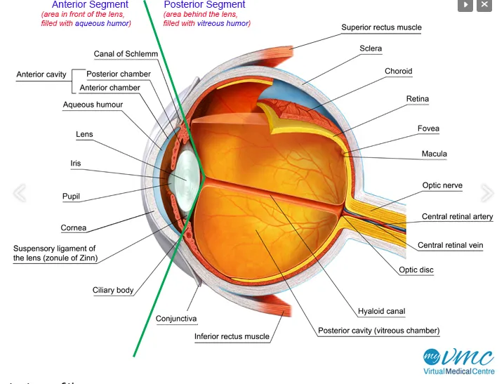
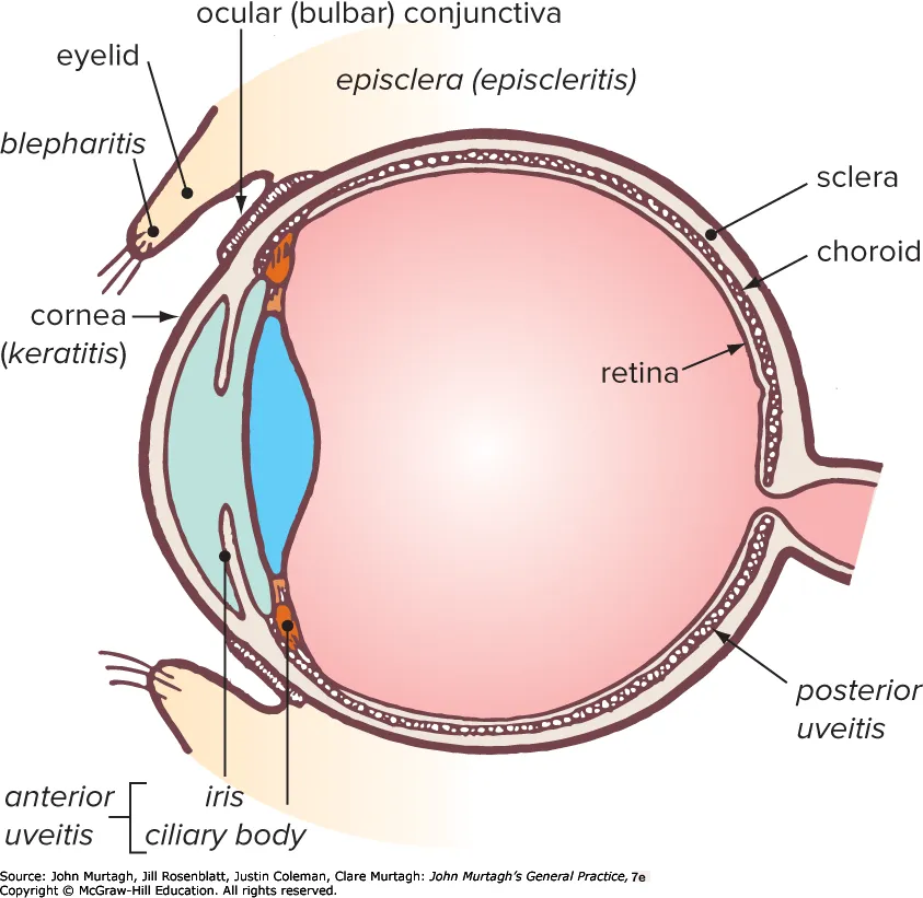
The eye is divided into an anterior segment (in front of the lens — cornea, anterior chamber, aqueous humour, iris/pupil, ciliary body and the lens’s suspensory ligament/zonule of Zinn, drained via the canal of Schlemm) and a posterior segment (behind the lens, filled with vitreous humour — choroid, retina, macula, optic nerve, central retinal artery/vein). Light reaching the retina passes through four normally transparent media — cornea, aqueous, lens, vitreous — in sequence; a hazy or absent red reflex localises the problem to an opacity somewhere along that path (commonly cataract or vitreous haemorrhage) rather than to retinal disease itself.
Normal fundus
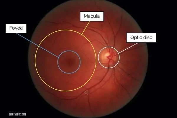
The key landmarks are the optic nerve head (optic disc) and the macula.
- The optic disc is found by tracing any blood vessel to the point of coalescence — branching vessels form an arrow pointing towards the disc. The disc is yellow-pink, sharply demarcated (more so on its temporal edge), and normally slightly taller than wide; its transverse diameter (~1.5mm) is the standard yardstick for describing the size and position of any other fundal finding (e.g. one-half disc diameter at 2 o’clock”). “Retinal arteries/arterioles are lighter, more orange-red, and visibly narrower than the paired veins/venules they run alongside — the colour difference reflects oxygenated versus deoxygenated blood seen through the transparent vessel wall, not a difference in the wall itself. The larger veins normally show a slight spontaneous pulsation; its presence reliably excludes a raised intracranial pressure sufficient to compress the vein, while its absence is a much less reliable sign on its own (a sizeable minority of normal eyes don’t pulsate).
- The macula lies temporal to the optic disc. Its central part, the fovea, is about the same diameter as the optic disc and appears darker due to additional pigment. It is the most light-sensitive part of the retina and the area of greatest visual acuity, so it is examined last (and only really assessable once the pupil is dilated) to avoid provoking pupilloconstriction and losing the view early.
No haemorrhage, exudate, or wrinkling should be visible in a normal fundus; any sharp elevation change that brings one area out of focus while the rest stays sharp is likewise abnormal.
Diabetic retinopathy
Diabetic retinopathy is a microangiopathy causing retinal small-vessel damage from
hyperglycaemia. Duration of diabetes and poor glycaemic control are the most important risk
factors; hypertension, hyperlipidaemia and smoking also contribute. See
diabetic-retinopathy.md for the full staging and management detail — this page covers the
fundoscopic appearance at each stage.
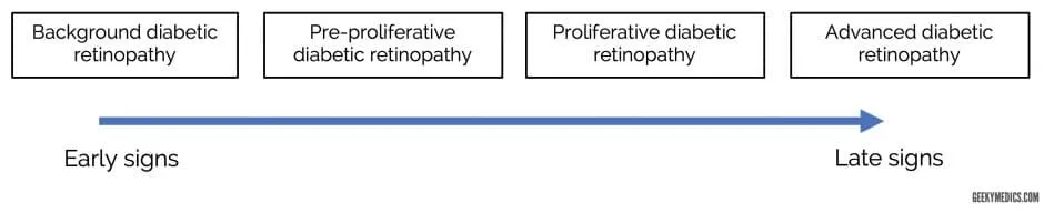
The formal Early Treatment Diabetic Retinopathy Study (ETDRS) classification, which underlies most contemporary staging, grades severity by the extent of haemorrhages and associated changes rather than by the informal groupings below alone: very mild non-proliferative disease shows microaneurysms only; mild non-proliferative disease adds a few haemorrhages/hard exudates; moderate disease shows retinal haemorrhages in 1-3 quadrants plus cotton wool spots; and severe non-proliferative disease meets the “4-2-1 rule” — severe haemorrhages in all four quadrants, venous beading in ≥2 quadrants, or moderate intraretinal microvascular abnormalities (IRMA) in ≥1 quadrant (any one criterion is sufficient; ≥2 criteria makes it “very severe”).
Background retinopathy — microaneurysms and dot/blot haemorrhages
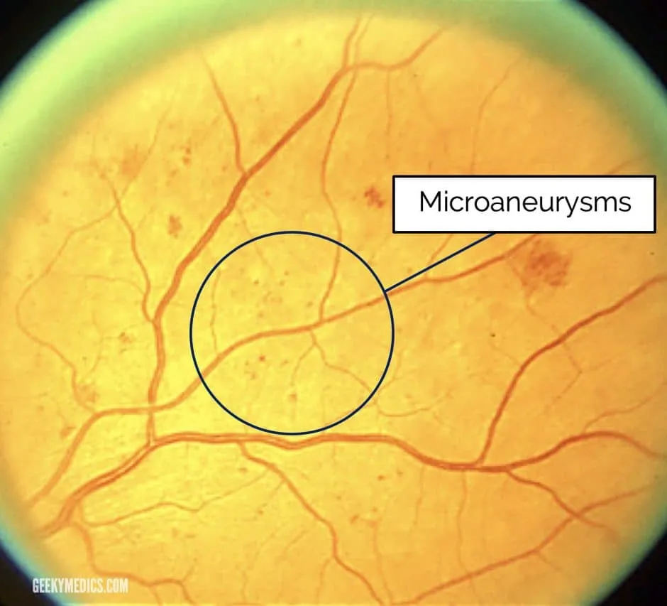
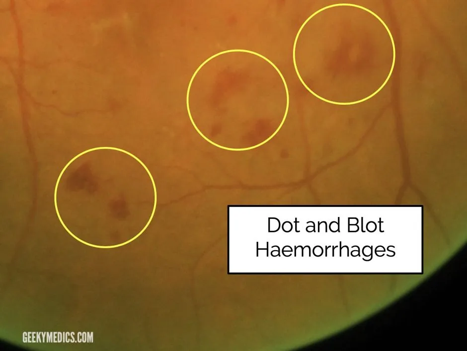
Microaneurysms and dot/blot haemorrhages (from bleeding capillaries in the middle retinal layers) can look similar when small; distinguishing them isn’t clinically important since both indicate pre-proliferative-stage disease. Microaneurysms are focal capillary-wall outpouchings; dot/blot haemorrhages sit deeper, in the inner nuclear and outer plexiform layers, and are typically larger with a more irregular margin.
Pre-proliferative retinopathy — cotton wool spots
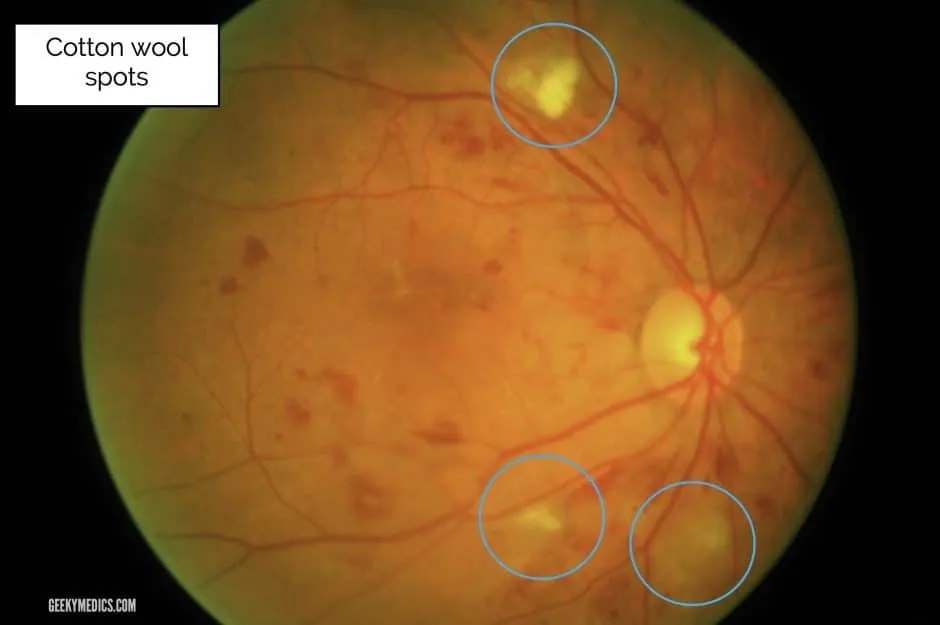
Retinal ischaemia marks progression from background to pre-proliferative disease. Cotton wool spots are small, fluffy, whitish superficial lesions — accumulations of dead nerve cells from ischaemic damage. Other pre-proliferative signs (venous changes, intraretinal microvascular anomalies/IRMA) are beyond undergraduate-level recognition but are what the formal “severe NPDR” grading above is actually counting.
Proliferative retinopathy
Insufficient retinal perfusion drives VEGF production and neovascularisation — new vessels at the disc (NVD) or elsewhere on the retina (NVE). High-risk” proliferative disease — the threshold for urgent laser treatment — is defined by NVD of at least a quarter to a third of a disc area, or any NVD/NVE accompanied by a vitreous or pre-retinal haemorrhage. “A Cochrane review of 59 prognostic studies found higher HbA1c and more advanced retinopathy at baseline are the most consistently supported risk factors for progressing to proliferative disease; renal impairment, younger age at diagnosis (in type 1 diabetes), and larger retinal venous calibre showed weaker, lower-certainty associations, while duration of diabetes, blood pressure, and cholesterol were not consistently associated with progression once retinopathy severity was accounted for.
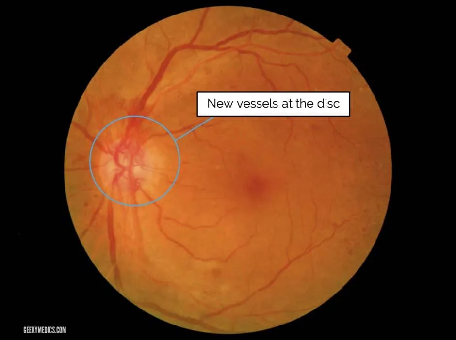
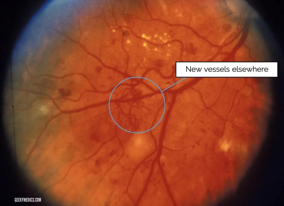
Advanced disease can produce recurrent vitreous haemorrhage (bleeding neovascular areas), tractional retinal detachment (fibrous bands from neovascularisation suspending the retina), and rubeosis (iris/drainage-angle neovascularisation causing secondary glaucoma).
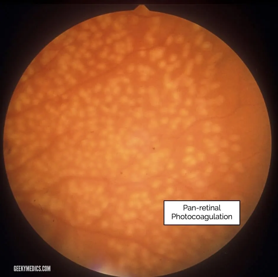
Pan-retinal photocoagulation (PRP) is the primary treatment for proliferative disease, reducing peripheral retinal oxygen demand and thus VEGF production. Seen clinically as clusters of laser burn marks.
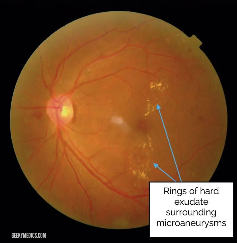
Diabetic maculopathy — exudates and/or macular oedema at the macula. Oedema is hard to visualise with a direct ophthalmoscope. Hard exudates are waxy yellow lesions with distinct margins, in clumps or rings around leaking microaneurysms (distinguished from “soft exudates” — the older term for cotton wool spots), and typically form at the border between oedematous and non-oedematous retina.
Hypertensive retinopathy
Acute BP elevation usually causes reversible retinal vasoconstriction; hypertensive crisis
can also cause optic disc oedema — disc oedema with elevated BP defines hypertensive crisis.
Prolonged severe hypertension causes exudative retinal changes (haemorrhages, hard exudates,
cotton wool spots). See hypertensive-retinopathy.md for full detail.
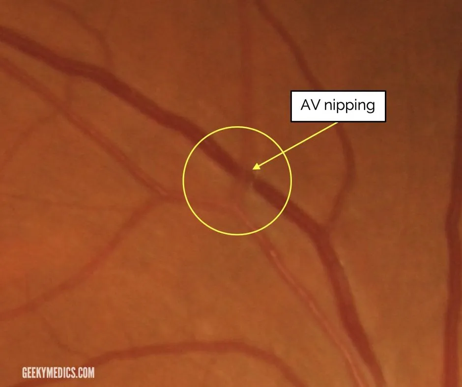
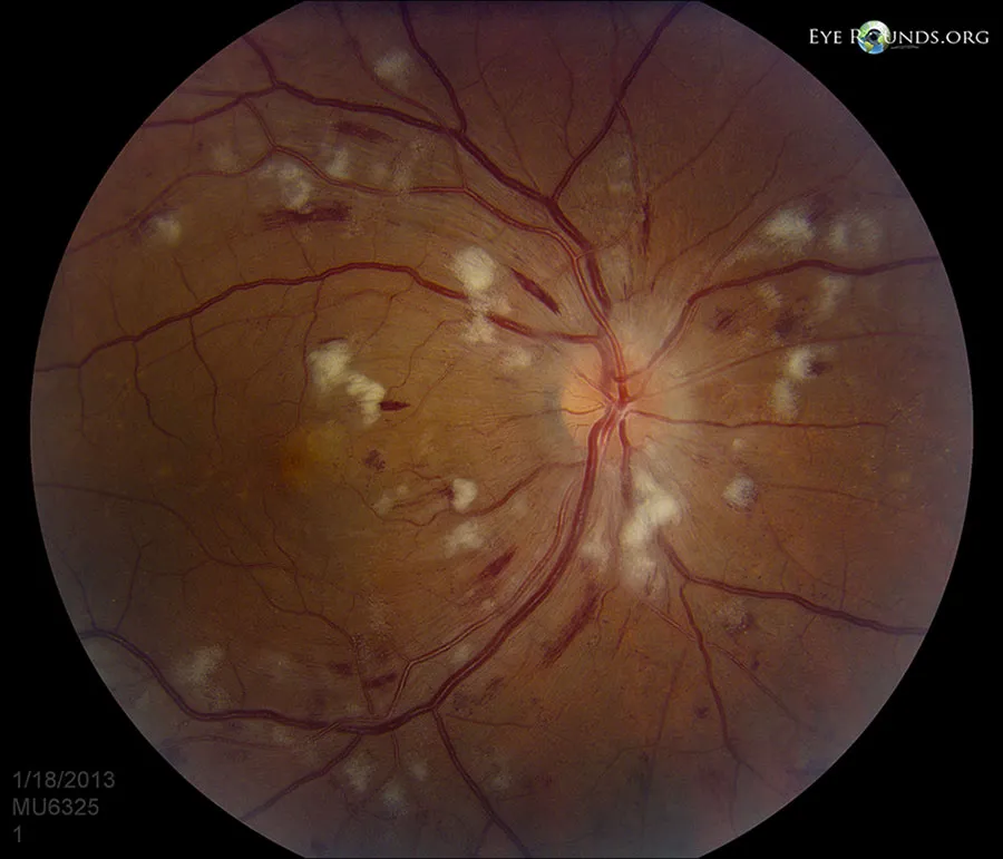
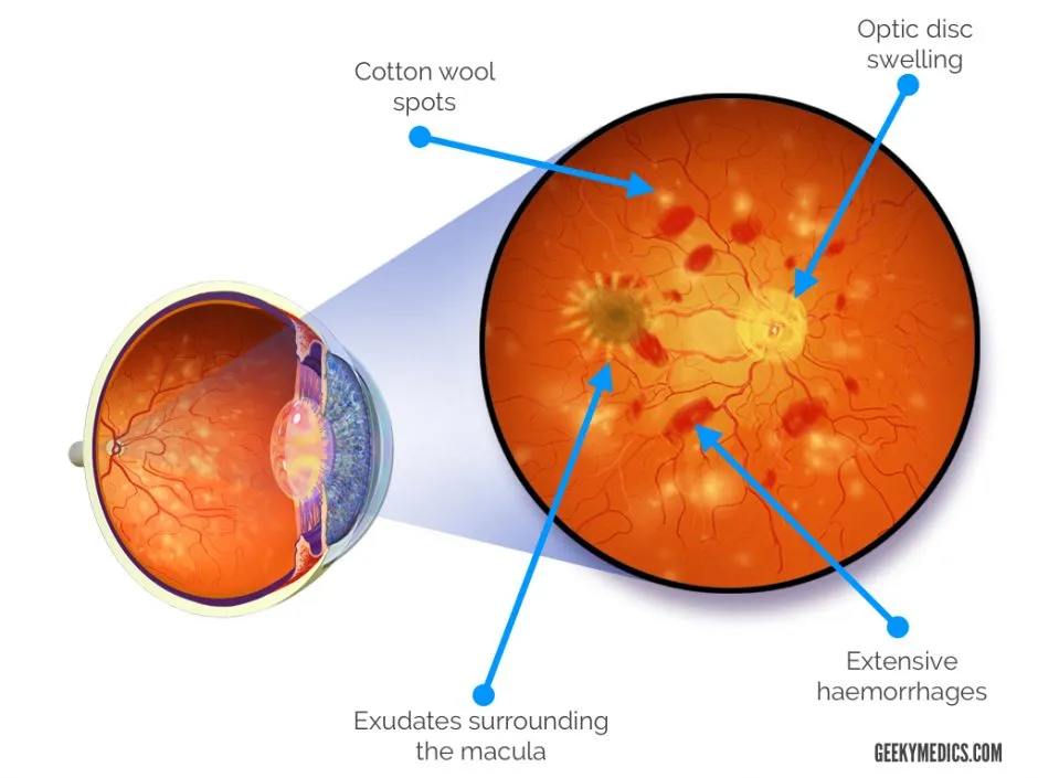
Chronic hypertension is usually asymptomatic early; late disease may show blurred vision and visual field deficits. The table below follows the same 4-group structure as the Keith-Wagener-Barker classification, the system still most widely quoted for correlating fundoscopic findings with hypertension severity and chronicity, though its prognostic value in isolation (independent of BP readings themselves) is debated. Chronic arteriolar wall thickening produces two further fundoscopic signs not shown in the table: a widened, accentuated central light reflex (“copper wiring”) in moderate disease, progressing to a duller, opaque “silver wiring” appearance in severe longstanding disease, where the vessel wall itself becomes visible rather than the blood column within it.
| Grade | Findings |
|---|---|
| 1 | Subtle generalised arteriolar narrowing |
| 2 | Focal arteriolar narrowing + venule compression at AV crossings (AV nipping) |
| 3 | Grade 2 + retinal haemorrhages, hard exudates, cotton wool spots |
| 4 (malignant hypertension) | Grade 3 + optic disc swelling, plus headaches, eye pain, reduced acuity, focal neurological deficits. Managed with antihypertensives + emergency admission |
Malignant hypertension is generally defined as a BP above ~200/120mmHg with this grade-4 bilateral retinopathy; untreated, mortality reaches up to 50% within 2 months and nearly 90% within a year, which is why grade-4 findings on fundoscopy are an admission trigger in their own right, independent of the absolute BP reading at the time.
Optic nerve pathology
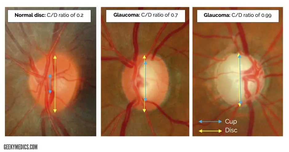
Glaucomatous disc changes — glaucoma is a progressive optic neuropathy with visual field loss, usually with raised intraocular pressure. Irreversible nerve fibre loss produces disc “cupping” — an increased vertical cup-to-disc ratio (normally the cup occupies less than about a third of the disc’s diameter; a cup-to-disc ratio ≥0.7, or a marked asymmetry between the two eyes, is a specific pointer towards glaucoma rather than a normally large physiological cup).
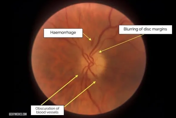
Papilloedema / optic disc swelling — papilloedema specifically means disc swelling from raised intracranial pressure; optic disc swelling” is the general term for any cause, including intracranial space-occupying lesions, optic neuritis (MS), malignant hypertension and uveitis (TB, sarcoidosis). Blurred disc margins ± small haemorrhages. “Findings progress with severity on the widely-used Frisén grading scale: grade 1 is blurring confined to the nasal disc border with a normal temporal margin; grade 2 adds circumferential blurring of both borders; grade 3 shows elevated, blurred margins obscuring one or more major vessels as they cross the disc; grade 4 shows total obscuration of a vessel segment; and grade 5 is complete obscuration of all disc vessels with the most pronounced elevation. As above, loss of spontaneous venous pulsation supports but does not confirm raised intracranial pressure, and its presence does not exclude early papilloedema (pulsation can be lost before visible disc swelling develops).
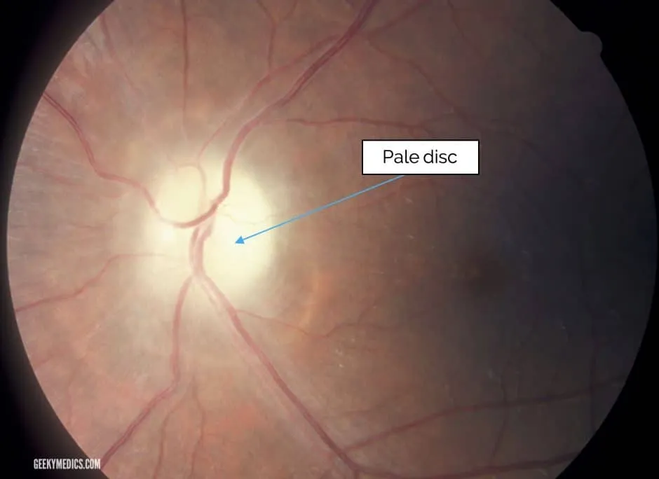
Optic nerve atrophy — death of nerve fibres producing a pale disc, usually taking 4-6 weeks to develop after the causative axonal injury. Primary atrophy: inflammation (optic neuritis), glaucoma, or retinal ischaemia, with an orderly pattern of axon loss that leaves the disc architecture intact — sharply demarcated margins, a chalky-white cupped disc, and attenuated vessels. Secondary atrophy: longstanding papilloedema (also optic neuritis or anterior ischaemic optic neuropathy), where disorderly axon loss with glial proliferation instead leaves the disc grey with indistinct margins and an obliterated cup. A third pattern, consecutive atrophy, follows disease of the inner retina or its blood supply (e.g. after central retinal artery occlusion, extensive photocoagulation, or retinitis pigmentosa) and looks different again — a waxy pale disc with markedly attenuated arteries but a normal margin and normal cup. Investigate any new optic atrophy with neuroimaging to exclude intracranial causes.
Vascular occlusive disease
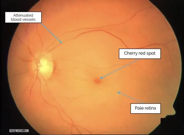
Central retinal artery occlusion (CRAO) — sudden, profound vision loss, most often from
embolism (e.g. stroke), occasionally vasculitis (giant cell arteritis). Classic “cherry-red
spot” — the intact foveal reflex standing out against the pale ischaemic retina. See
retinal-vascular-occlusions.md — this is now managed as an acute ischaemic stroke
equivalent.
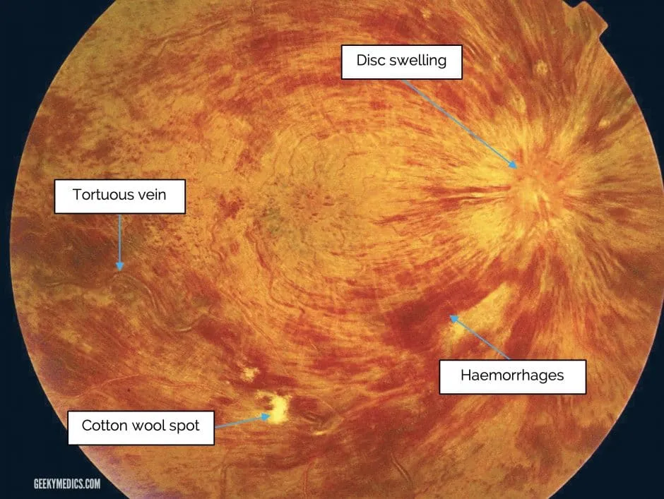
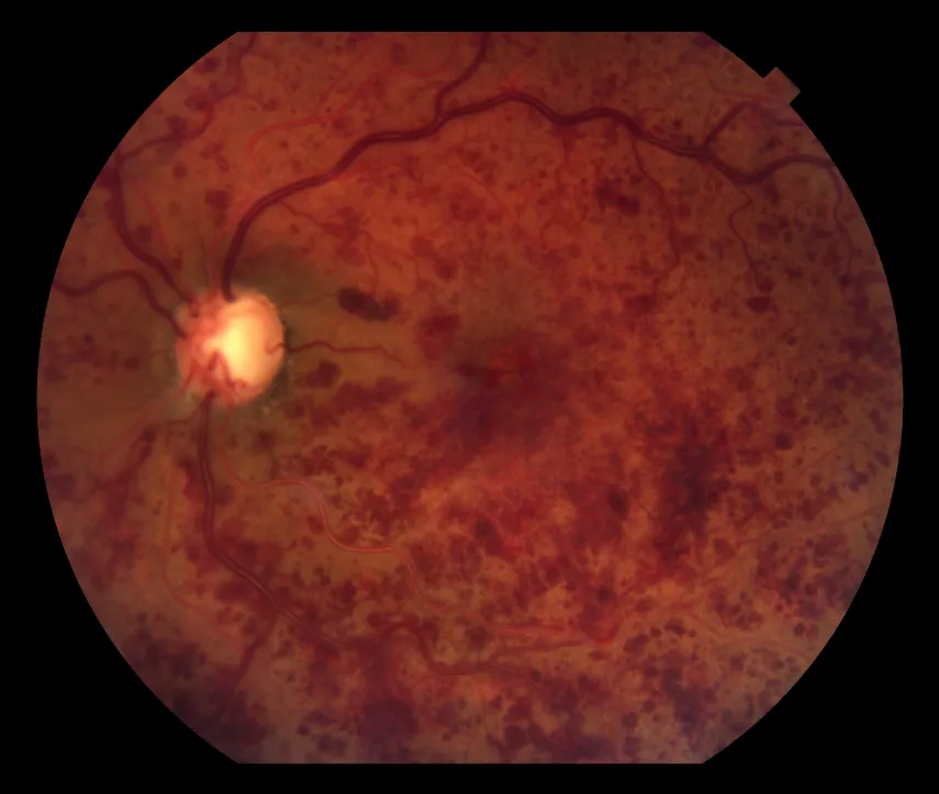
Central retinal vein occlusion (CRVO) — painless vision loss, usually sudden but may progress over days to weeks. Risk factors: hypertension and age (major, ~90% of cases occur over 50), glaucoma, diabetes, raised blood viscosity. Occurs from atherosclerotic thickening of the central retinal artery compressing the vein at their shared crossing point, causing retinal hypoxia and extravasation of blood. The classic fundoscopic description is a “blood and thunder” fundus — extensive haemorrhages through all four quadrants — with tortuous, engorged retinal veins, cotton wool spots and disc swelling. Non-ischaemic (perfused) CRVO accounts for around 70% of cases and has milder findings and a better visual prognosis than ischaemic (non-perfused) CRVO, which shows more marked venous dilation and oedema and is defined by at least 10 areas of capillary non-perfusion on angiography.
Prognosis correlates with presenting vision: ≥6/12 favours spontaneous improvement; 6/60
makes improvement unlikely with a risk of further deterioration. Treatment: intravitreal
anti-VEGF or dexamethasone, laser photocoagulation. See retinal-vascular-occlusions.md.
Macular pathology
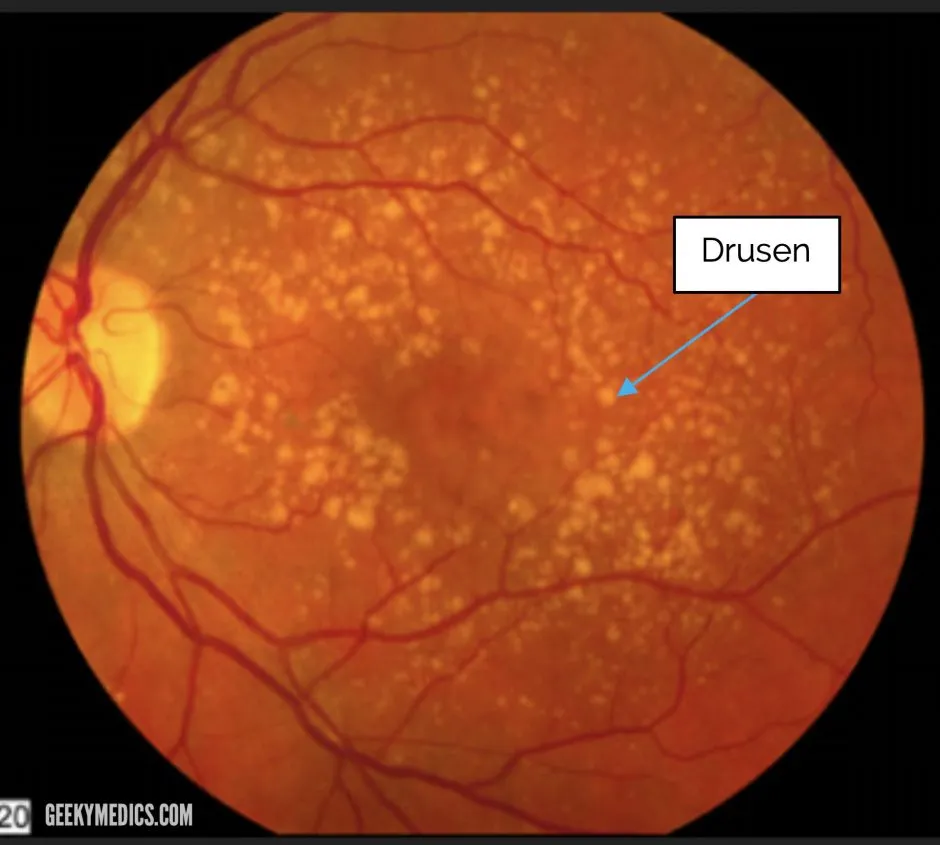
Age-related macular degeneration (AMD) — a degenerative macular disorder and the most
common cause of irreversible blindness in the elderly. ~90% of cases are the “dry” form (no
exudation or neovascularisation). Early sign: yellow-white drusen deposits. See
macular-degeneration.md for current management (AREDS2 supplementation, anti-VEGF for wet
AMD).
Source: https://geekymedics.com/fundoscopic-appearances-of-retinal-pathologies/