Diabetic Retinopathy
Written 20th July 2020 - Last Update 20th August 2026
Diabetic Retinopathy
A microangiopathy causing damage to small retinal blood vessels from hyperglycaemia. It is the most common microvascular complication of diabetes and the leading cause of vision loss in working-age adults, affecting an estimated 77% of people with type 1 diabetes and 25% of people with type 2 diabetes over their lifetime; around a quarter to a third of those affected go on to develop vision-threatening diabetic macular oedema.
Risk factors for developing diabetic retinopathy at all: duration of diabetes and poor glycaemic control (HbA1c) are the most important; hypertension, hyperlipidaemia, obesity, nephropathy and smoking also contribute, along with non-modifiable factors (puberty, pregnancy).
Risk factors specifically for progressing to proliferative disease (PDR) are narrower. A 2023 Cochrane review of 59 prognostic studies (87 articles, ~113,000 participants) found higher HbA1c and more advanced baseline retinopathy severity were the most consistently supported, moderate-certainty risk factors for progression to PDR; renal impairment, younger age at diagnosis (in type 1 diabetes), higher triglycerides and larger retinal venous calibre showed weaker, low-certainty associations. Notably, duration of diabetes, blood pressure, cholesterol, BMI, ethnicity and smoking were not found to be independently associated with progression to PDR once baseline retinopathy severity was accounted for — a different question from the risk factors for developing retinopathy in the first place.
Staging
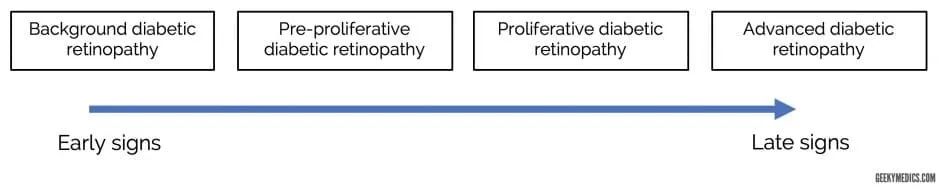
The Early Treatment Diabetic Retinopathy Study (ETDRS) classification underlies most contemporary staging, grading non-proliferative disease by the extent of haemorrhages and associated changes: very mild NPDR shows microaneurysms only; mild NPDR adds a few haemorrhages/hard exudates; moderate NPDR shows retinal haemorrhages in 1–3 quadrants plus cotton wool spots; severe NPDR meets the “4-2-1 rule” — severe haemorrhages in all four quadrants, venous beading in ≥2 quadrants, or moderate intraretinal microvascular abnormalities (IRMA) in ≥1 quadrant (any one criterion is sufficient); very severe NPDR meets two or more of those criteria.
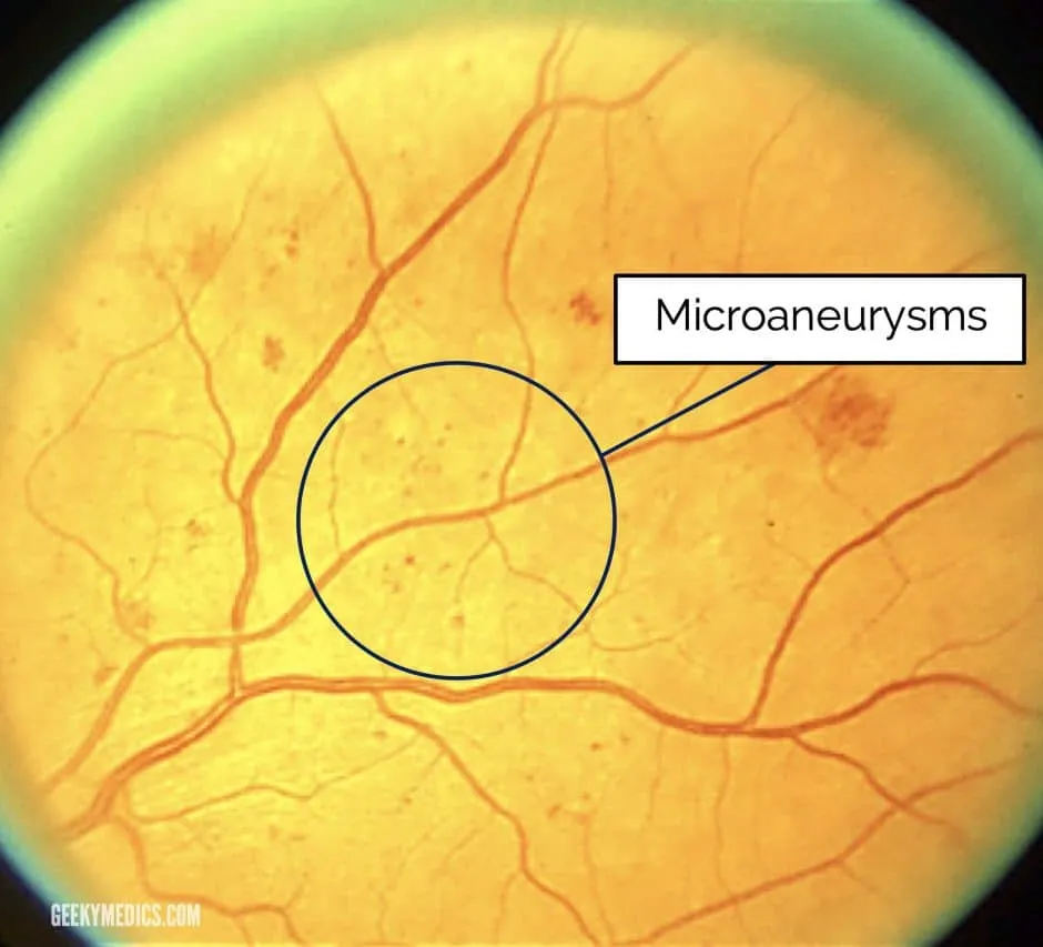
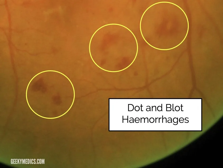
Background retinopathy — microaneurysms and dot/blot haemorrhages
Dot and blot haemorrhages arise from bleeding capillaries in the middle retinal layers and can look like microaneurysms when small — distinguishing the two isn’t clinically important since both belong to the pre-proliferative spectrum.
Pre-proliferative retinopathy
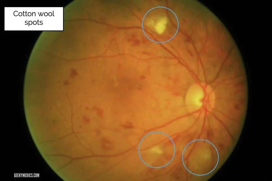
Retinal ischaemia marks progression from background to pre-proliferative disease. Cotton wool spots — small, fluffy, whitish superficial lesions — are accumulations of dead nerve cells from ischaemic damage. Other pre-proliferative signs (venous changes, intraretinal microvascular anomalies/IRMA) are beyond undergraduate-level recognition but are what the formal “severe NPDR” grading above is actually counting.
Proliferative retinopathy
Insufficient retinal perfusion drives VEGF production and neovascularisation — new vessels at the disc (NVD) or elsewhere (NVE). High-risk proliferative disease — the threshold for urgent laser treatment — is defined by NVD of at least a quarter to a third of a disc area, or any NVD/NVE accompanied by a vitreous or pre-retinal haemorrhage.
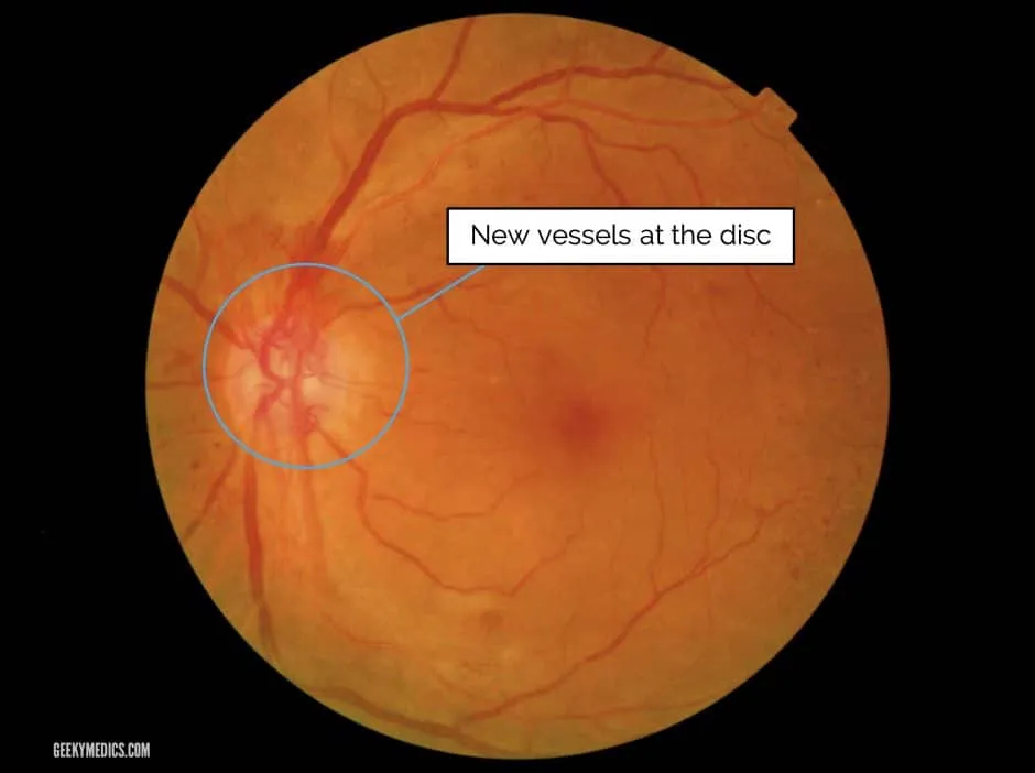
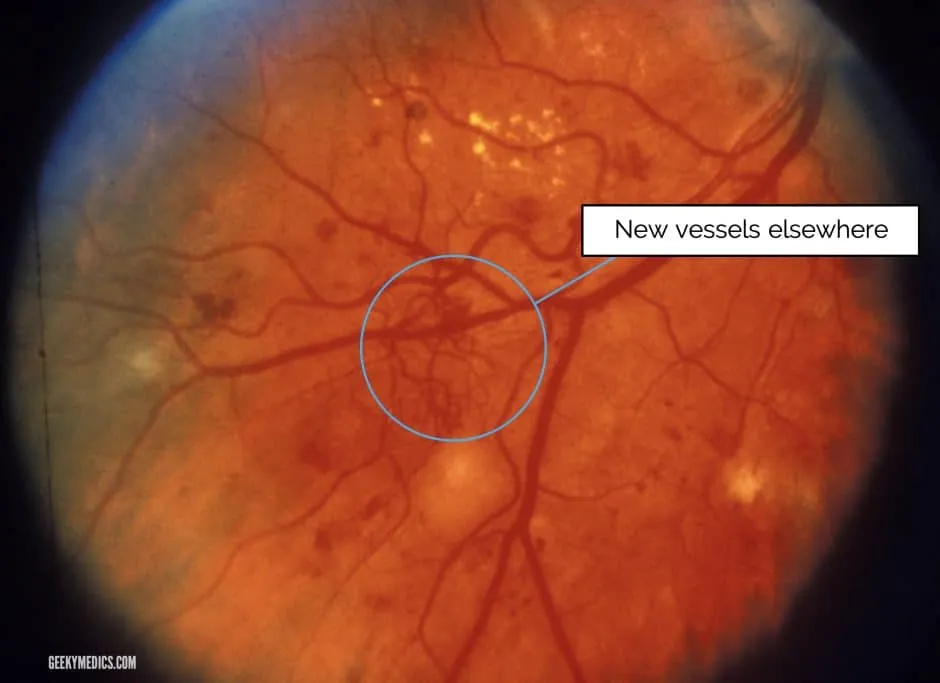
Advanced disease can produce recurrent vitreous haemorrhage (bleeding neovascular areas), tractional retinal detachment (fibrous bands from neovascularisation suspending the retina), and rubeosis (iris/drainage-angle neovascularisation causing secondary glaucoma).
Screening and follow-up
Follow-up interval is set by NPDR severity: very mild NPDR is reviewed annually; mild-to- moderate NPDR every 6–12 months (16% of mild and 23% of moderate NPDR progress to PDR within 4 years); severe-to-very-severe NPDR needs close review every 2–4 months (50% of severe and 75% of very severe NPDR progress to PDR within 1 year). Strict glycaemic control (HbA1c <7%) and management of associated hypertension, dyslipidaemia and nephropathy remain the foundation of slowing progression at every stage.
Treatment
Pan-retinal photocoagulation (PRP) is the traditional primary treatment for proliferative disease — laser burns to the peripheral retina reduce oxygen demand and thus VEGF production. Treatment is tiered by risk: non-high-risk PDR without macular oedema is managed with close 2–4-monthly review, moving to PRP once high-risk features develop; high-risk PDR without macular oedema is treated with PRP; PDR with concurrent macular oedema is managed with combined anti-VEGF injections and PRP; and where complete PRP isn’t possible (e.g. media opacity), cryopexy or vitrectomy are the alternatives.
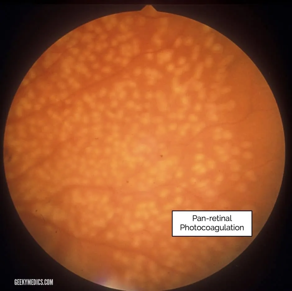
Anti-VEGF injections (e.g. aflibercept, ranibizumab, bevacizumab) are now widely used for diabetic macular oedema, and increasingly for proliferative disease alongside or instead of PRP — a large randomised trial (DRCR.net Protocol S) found intravitreal ranibizumab non-inferior to PRP for PDR, though it trades a one-off laser session for repeated injections and closer follow-up. (Specific dosing for these agents is not detailed here — cross-check against eTG/AMH before prescribing, as the published trial doses are from US-facing references not yet verified against Australian guidelines.)
Vitrectomy is indicated for non-clearing vitreous haemorrhage, tractional retinal detachment threatening or involving the macula, combined tractional–rhegmatogenous detachment, anterior-segment neovascularisation obscuring the view of the posterior segment, or dense epiretinal membrane/vitreomacular traction.
Diabetic maculopathy
Exudates and/or macular oedema at the macula — oedema is hard to visualise with a direct ophthalmoscope. Hard exudates are waxy yellow lesions with distinct margins in clumps or rings around leaking microaneurysms (distinguished from “soft exudates” — the older term for cotton wool spots).
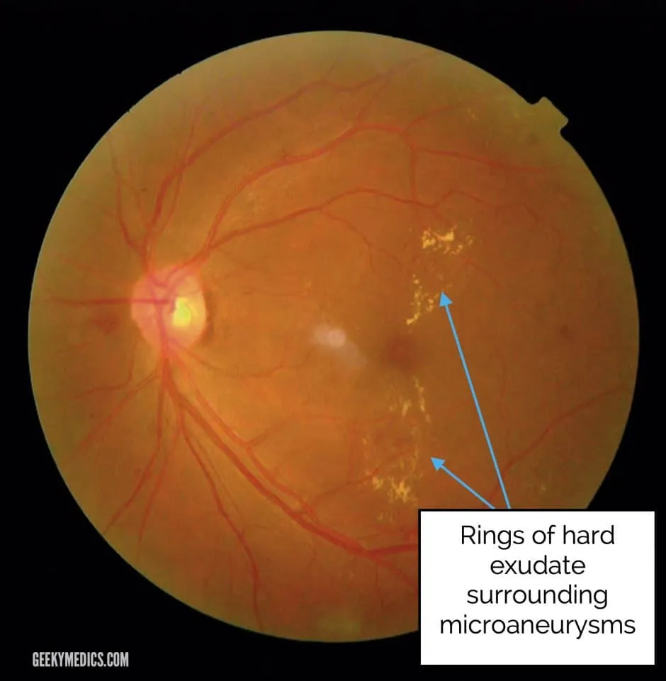
Clinically significant macular oedema (CSME), the ETDRS treatment threshold, is defined as any of: retinal thickening within 500 microns of the foveal centre; hard exudates within 500 microns of the centre if associated with adjacent retinal thickening; or an area of retinal thickening at least one disc area in size, any part of which lies within one disc diameter of the centre. Anti-VEGF injections are now first-line for centre-involving macular oedema; focal/grid laser remains the treatment of choice for non-centre-involving oedema — ETDRS showed laser halts progression of moderate vision loss but does not itself improve visual acuity. Intravitreal corticosteroid implants are reserved for eyes not responding to anti-VEGF therapy.
Diabetic retinopathy in pregnancy
Progression occurs in both type 1 and type 2 diabetes, especially in the second/third trimesters (14% prevalence in type 2 diabetes during pregnancy). Risk factors for worsening: baseline retinopathy severity, HbA1c control, diabetes duration, and hypertension. Regression and spontaneous visual recovery often occur postpartum. Gestational diabetes carries a very small (<1%) retinopathy risk — ophthalmic exam isn’t routinely needed for gestational diabetes alone.
RANZCO recommends: ocular exam before conception and again in the first trimester for women with pre-existing diabetes, with referral to ophthalmology within 4 weeks of that initial exam. Intravitreal anti-VEGF safety is not established in pregnancy and should be avoided — intravitreal triamcinolone is the preferred alternative if treatment for diabetic macular oedema is needed. Non-clearing vitreous haemorrhage or tractional retinal detachment may need surgical intervention regardless of pregnancy.