Primary Open-Angle & Normal-Tension Glaucoma
Written 9th July 2020 - Last Update 23rd July 2026
Primary Open-Angle & Normal-Tension Glaucoma
Glaucoma is best understood as a progressive optic neuropathy, with intraocular pressure (IOP) a modifiable risk factor rather than the disease itself. It is the second leading cause of irreversible blindness in Australia in people over 55, and 50–60% of Australian cases remain undiagnosed — the economic cost was $4.3 billion in 2025 (up from $1.9 billion in 2005).
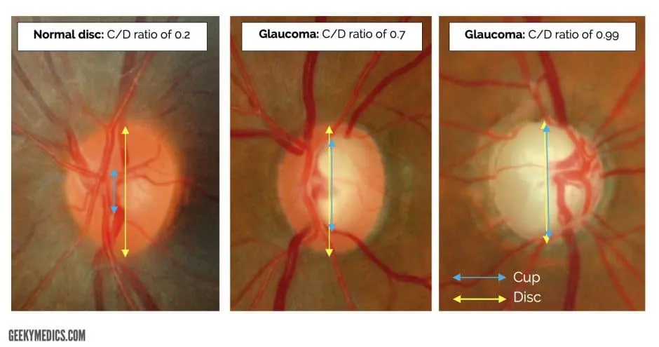
Glaucoma causes irreversible retinal nerve fibre loss, seen as optic disc “cupping” — an increased vertical cup-to-disc ratio.
Classification
| Type | Presentation | Urgency |
|---|---|---|
| Primary open-angle glaucoma (POAG) | Insidious, asymptomatic, disc cupping ± raised IOP | Routine ophthalmology |
| Normal-tension glaucoma (NTG) | Glaucomatous disc/field change with normal IOP | Routine ophthalmology |
| Primary angle-closure glaucoma (chronic) | Progressive peripheral field loss, shallow anterior chamber | Routine ophthalmology |
| Secondary glaucomas | Steroid-induced, pseudo-exfoliative, uveitic, pigmentary, traumatic, neovascular | As per underlying cause |
See acute-glaucoma.md for the acute angle-closure emergency.
Epidemiology and risk factors
- Commonest cause of irreversible blindness in middle age; ~1 in 8 people over 80
- Family history: a first-degree relative confers roughly a 2.5-fold increased risk
- Age: risk rises ~7% per year; those aged 60–69 have roughly triple the risk of those 40–49
- Ethnicity: African and Asian ancestry carry disproportionately higher risk than European ancestry
- Other risk factors: diabetes, myopia (also raises hypermetropia-related angle-closure risk), long-term corticosteroid use, migraine/peripheral vasospasm, abnormal blood pressure, prior eye trauma
- Ocular hypertension remains the single strongest modifiable risk factor — risk rises with each 1 mmHg increment
- Male sex is associated with higher POAG risk; angle-closure glaucoma is more frequent in females
Presentation
- Early: asymptomatic
- Late: insidious peripheral visual field loss; central vision preserved until advanced
Screening (RACGP Red Book-style risk-based approach)
Screen those at higher risk (which is most people over 50):
- First-degree relative with glaucoma → refer for ocular examination 5–10 years earlier than the relative’s age of onset
- Caucasian/Asian patients ≥50 years; patients of African descent ≥40 years
- Any age with: diabetes, myopia, long-term steroid use, migraine/peripheral vasospasm, abnormal BP, prior eye trauma
Refer for optic nerve head examination (ophthalmoscopy), tonometry, and visual field assessment (perimetry) — by an ophthalmologist or optometrist. Follow-up frequency is individualised; no fixed consensus interval exists.
GP-relevant referral thresholds (2026 AJGP framework)
| Finding | Likely condition | Action |
|---|---|---|
| Sudden VA drop, new field loss, fresh optic-disc (Drance) haemorrhage, or vertical cup-to-disc ratio ≥0.8/asymmetry ≥0.3 | Rapidly progressive open-angle glaucoma | Urgent referral ≤1 week |
| IOP ≥35 mmHg (even asymptomatic), or IOP 25–34 mmHg with a suspicious disc or reproducible visual field defect | Sight-threatening ocular hypertension | Urgent referral ≤48 hours |
| IOP 22–25 mmHg without other risk factors; or a high-risk patient (age >60, FHx, diabetes, steroid use) even with normal IOP | Possible early POAG/NTG | Routine referral 1–3 months |
Formal visual field testing and OCT imaging (used to confirm progression) are outside GP scope — performed by optometrists/ophthalmologists. Direct ophthalmoscopy in an undilated pupil gives only a rough cup-to-disc estimate; interpret cautiously, but marked inter-eye asymmetry or a clearly enlarged cup still warrants referral. Tonometry is not routinely available in general practice and is not required to trigger a referral decision.
Management
Managed by ophthalmology. Treatment cannot restore lost vision but slows disease progression — lifelong therapy aimed at reducing IOP:
| Line | Class | Mechanism | Example | Notable adverse effects |
|---|---|---|---|---|
| First | Prostaglandin analogues | ↑ uveoscleral outflow | Latanoprost | Iris/lash pigmentation, local irritation |
| Second | β-blockers | ↓ aqueous production | Timolol | Systemic: bradycardia, hypotension, bronchospasm (caution in cardiac/respiratory disease) |
| Third | α-agonists | ↓ production + ↑ outflow | — | Fatigue, dry mouth, drowsiness, delayed allergic conjunctivitis |
| Third | Carbonic anhydrase inhibitors | ↓ production | — | Paraesthesia, GI upset, electrolyte disturbance, metabolic acidosis (oral agents especially) |
PBS note: the 60-day dispensing model now allows up to 12 months’ supply for some glaucoma medications, alongside typically annual ophthalmology review — reinforcing drop technique, monitoring for the adverse effects above, and timely script renewal are the key GP contributions to preventing avoidable progression.
The GP’s coordinating role
- Coordinate and manage — ensure continuity of topical therapy, address comorbidities, arrange timely review
- Refer rather than defer — any suspected glaucoma (elevated IOP, suspicious disc, relevant symptoms/risk factors) goes to an eye care specialist for baseline assessment; go directly to ophthalmology when red-flag criteria (table above) are met
- Shared records — copy referral correspondence to both optometrist and ophthalmologist where a shared-care model is in place