Acute Angle-Closure Glaucoma
Written 15th July 2020 - Last Update 23rd July 2026
Acute Angle-Closure Glaucoma
A sudden rise in intraocular pressure (IOP), typically >30–50 mmHg, from apposition of the iris against the trabecular meshwork blocking aqueous outflow.
Risk factors
Long-sightedness (hypermetropia), female sex, increasing age.
Presentation
- Sudden-onset painful red eye
- Haloes around lights
- Nausea and vomiting
- Headache
- Usually occurs in low light, when the pupil is dilated
- Bilateral involvement in an acute attack is rare
Examination
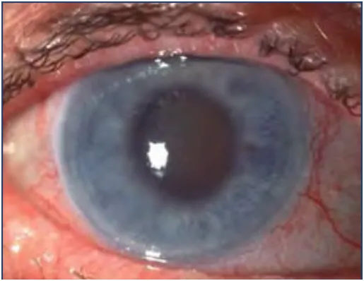
- Hazy/steamy cornea (corneal oedema)
- Semi-dilated, fixed pupil
- Eye tense to palpation
- Conjunctival injection
Intermittent (prodromal) angle-closure attacks
A related but distinct presentation: transient brow ache or blurred vision specifically in dim light, haloes, and episodes of nausea/vomiting, with a shallow anterior chamber on oblique flashlight testing. Still needs same-day ophthalmology/ED referral — don’t mistake it for migraine because it resolves.
Investigations
Tonometry (if available) — IOP often ≥30 mmHg. The oblique flashlight test is a simple in-clinic screen for a shallow anterior chamber/angle-closure risk. Tonometry is not required to trigger referral if the clinical picture is convincing.
Management
Refer emergently — same-day ophthalmology or emergency department. IOP must be monitored by ophthalmology to confirm treatment is working.
Treatment aims to lower IOP (reduce aqueous production, constrict the pupil) and prevent iris–trabecular meshwork adhesion:
- IV acetazolamide
- Pilocarpine eye drops — constrict the pupil, prevent adhesion, improve outflow
- If no contraindication, a topical β-blocker (e.g. timolol) can be given while arranging transfer, alongside oral acetazolamide
Definitive treatment: laser peripheral iridotomy (or surgical iridectomy) — creates a hole in the peripheral iris to allow drainage and bypass the pupillary block.
Adjuncts: analgesia, antiemetics.