Abnormal Eye Movements (Diplopia)
Written 10th June 2020 - Last Update 23rd July 2026
Abnormal Eye Movements (Diplopia)
Usually acute in onset and distressing. Establish first whether it is monocular (problem within the eye itself — e.g. cataract, refractive error, corneal irregularity) or binocular (problem with the extraocular muscles/their innervation — resolves when either eye is covered).
Differential diagnosis
Common:
- Ocular cranial nerve palsies (III/IV/VI)
- CVA/TIA
- Ophthalmoplegic migraine
- Alcohol/benzodiazepines
- Monocular causes: cataract, refractive error, corneal pathology
By category:
| Category | Causes |
|---|---|
| Vascular | Stroke/TIA, microvascular ischaemia (diabetes, hypertension, atherosclerosis), aneurysm, carotid-cavernous fistula |
| Infective | Intraocular abscess, syphilis, mastoiditis, meningitis, herpes zoster |
| Neoplastic | Slow-growing tumour, raised ICP, skull-base tumours, infiltrative disease (e.g. leukaemia) |
| Autoimmune | Giant cell arteritis, multiple sclerosis, myasthenia gravis, orbital myositis, sarcoidosis, vasculitides, Guillain-Barré (a late sign) |
| Traumatic | Orbital wall fracture |
| Endocrine | Thyroid eye disease — see thyroid-eye-disease.md |
| Congenital | Decompensated childhood strabismus — see strabismus.md |
Malignancy should be considered in any patient with new-onset diplopia and a history of cancer — arrange prompt neuroimaging.
Giant cell arteritis — do not miss this
GCA must be excluded in any patient aged over 50 with diplopia. Diplopia occurs in up to 19% of GCA patients and can precede severe, potentially bilateral visual loss — treating the diplopia-causing episode promptly can prevent that outcome. Diplopia in GCA may be central (ischaemia of cranial nerve nuclei/supranuclear gaze centres) or peripheral (ischaemic cranial nerve or extraocular muscle insult) — sixth nerve palsy is a recognised isolated presentation, not just amaurosis fugax or vision loss.
- Systemic clues: constitutional symptoms (fever, night sweats, weight loss, fatigue, myalgia) in up to 50%; headache in ~60%; scalp tenderness, jaw claudication, temporal artery tenderness
- Investigate: ESR/CRP/platelets (elevated in most GCA, individual sensitivity 65–71%); temporal artery ultrasound (a “halo sign” — less invasive than biopsy, though biopsy remains gold standard: 77% vs 54% sensitivity, 100% vs 81% specificity)
- Do not delay steroids awaiting investigation results if clinical suspicion is high — refer urgently
- Treatment: IV methylprednisolone 3–5 days then a prolonged oral prednisolone taper over 1–2 years; tocilizumab (anti-IL-6) is increasingly used as a steroid-sparing agent with superior sustained-remission outcomes vs prednisolone alone
Examination
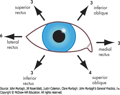
- Test the direction of gaze in which diplopia occurs, both monocularly (to exclude monocular diplopia) and binocularly
- Diplopia on lateral gaze → 6th nerve (abducens) palsy
- Diplopia on downward gaze (eye deviates upward) → 4th nerve (trochlear) palsy
- Eye deviated down and out → 3rd nerve (oculomotor) palsy
Investigations
- Screen for diabetes
- ESR/CRP for GCA (as above)
- TFTs for hyperthyroidism/thyroid eye disease
- Lipids for microvascular disease (more common with isolated abducens/CN VI palsies)
- MRI (preferred) or CT brain if MRI unavailable — for microvascular disease, TIA/CVA, and tumour identification
Management
- Discuss with ophthalmology or neurology
- Microvascular 6th nerve palsies typically resolve spontaneously in 3–6 months
- Surgery or botulinum toxin for a permanent deviation
- Patching one eye or an occluder gives symptomatic relief while a cause is identified/
treated — see
vision-and-fitness-to-drive.mdfor the driving implications of diplopia