Tuberous Sclerosis
Written 30th May 2020 - Last Update 28th August 2026
Tuberous Sclerosis
Autosomal dominant. Caused by a mutation in TSC1 (chromosome 9, hamartin) or TSC2 (chromosome 16, tuberin) — both normally restrain the mTOR pathway, so loss-of-function drives hamartoma growth across multiple organs.
- About a third of cases are inherited (50% risk to each child); the rest are sporadic, usually TSC2
- Prevalence ~1 in 20,000; median age at presentation ~7 months
Clinical features
Neurological
- Epilepsy in 70–90% of cases — may precede the skin findings, usually starting before age 3
- Cortical/subcortical tubers and subependymal nodules on imaging
- Subependymal giant cell astrocytoma (SEGA) in 10–20%
- Developmental delay, intellectual disability and behavioural problems (including autism spectrum disorder) in up to 90%
Skin (present in most patients and often the presenting clue)
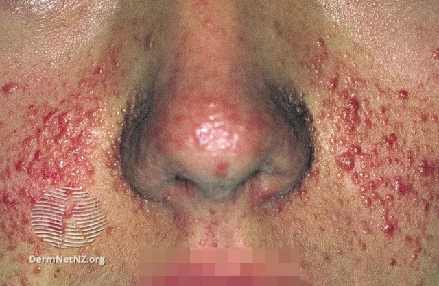
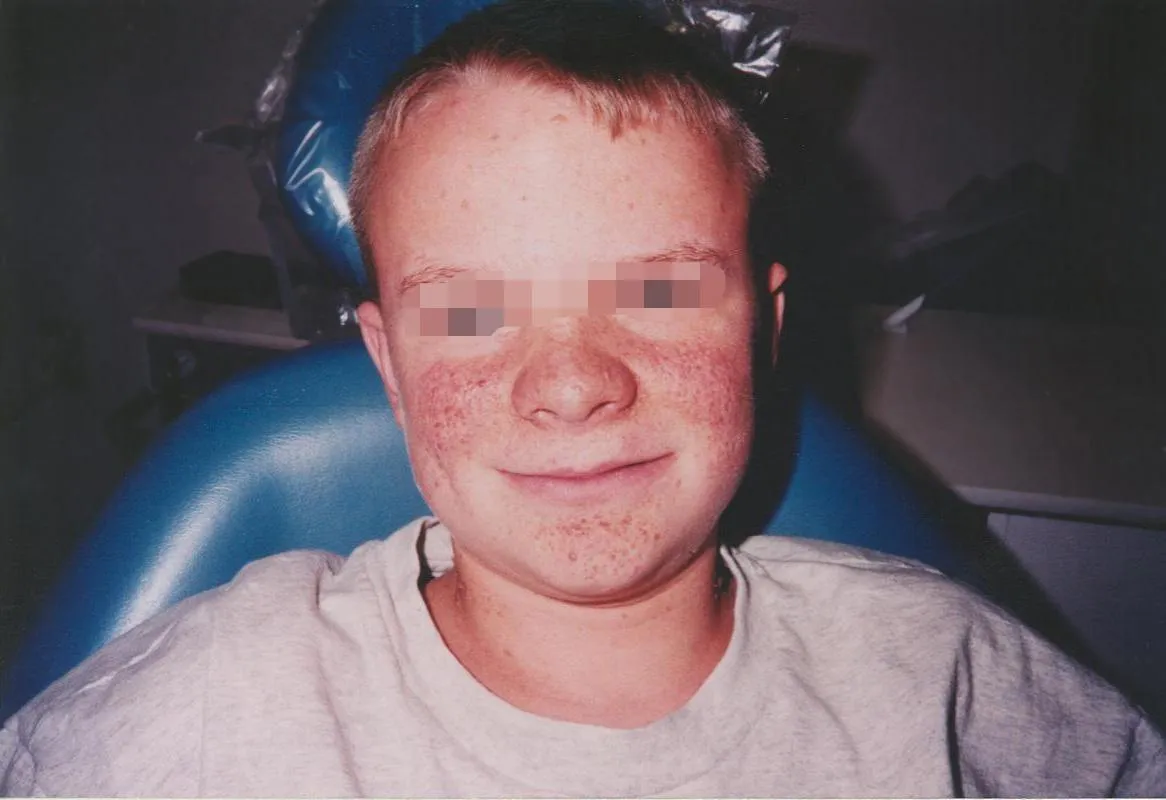
- Facial angiofibromas — the most obvious sign: pink/red papules on the cheeks and nose in a butterfly distribution, usually appearing between ages 3–10 and increasing through adolescence
- Ash-leaf (hypomelanotic) macules — ovoid white macules, often present from birth; three or more at birth is itself suggestive of the diagnosis
- Shagreen patches — flesh-coloured, orange-peel-textured connective tissue naevi, usually on the lower back
- Periungual/ungual fibromas — smooth flesh-coloured lumps emerging from the nail fold, more common on the toes; may show splinter haemorrhages
- Fibrous cephalic plaques and gingival fibromas also occur
Other organ systems
- Renal — angiomyolipomas (up to 75% by age 10–11); risk of pain, haemorrhage and, less commonly, renal cell carcinoma or polycystic kidney disease
- Cardiac — rhabdomyomas, usually present prenatally/in infancy and regressing spontaneously in childhood; can rarely cause neonatal heart failure
- Pulmonary — lymphangioleiomyomatosis (LAM), almost exclusively in women of reproductive age (median diagnosis age ~35)
- Ophthalmic — retinal astrocytic hamartomas
Diagnosis
2012 International TSC Consensus criteria: 2 major features, or 1 major + 2 minor, is diagnostic. A TSC1/TSC2 mutation is independently diagnostic on its own — but ~15% of clinically diagnosed patients have no identifiable mutation.
- Major: ≥2 hypomelanotic macules (≥5 mm), ≥2 angiofibromas or a fibrous cephalic plaque, ≥2 ungual fibromas, a shagreen patch, multiple retinal hamartomas, cortical dysplasia, subependymal nodules, SEGA, cardiac rhabdomyoma, LAM, multiple renal angiomyolipomas
- Minor: confetti skin lesions, >3 dental enamel pits, ≥2 intraoral fibromas, a retinal achromic patch, multiple renal cysts, non-renal hamartomas
MRI is the most sensitive imaging for tubers, subependymal nodules and SEGA. A Wood’s lamp helps pick up subtle hypomelanotic macules on exam.
Management
- Anti-epileptic drugs for seizure control — vigabatrin is particularly used for infantile spasms; around a third of patients have drug-refractory epilepsy and may need surgery
- mTOR inhibitors (sirolimus, everolimus) are first-line for asymptomatic/growing SEGA and for renal angiomyolipomas 3–6 cm — everolimus is TGA-approved for TSC-associated SEGA and refractory epilepsy; larger or symptomatic (bleeding) angiomyolipomas are managed with arterial embolisation or nephron-sparing surgery
- Topical rapamycin (sirolimus) is effective for facial angiofibromas, more so in children than adults; laser therapy is an alternative for cosmetic concerns
- Surgical resection for acute symptomatic SEGA (e.g. with hydrocephalus)
- Surveillance: annual clinical review, periodic brain MRI (tuber/SEGA monitoring), renal imaging for angiomyolipoma growth, and — specific to women — LAM screening (HRCT chest) from age 18, repeated every 5–7 years until menopause; women should be counselled that oestrogen exposure (including the combined pill) can worsen LAM
- Refer to neurology; multidisciplinary surveillance (nephrology, cardiology, ophthalmology, dermatology, and a specialised TSC service where available)