Tennis Elbow / Epicondylitis & Olecranon Bursitis
Written 13th February 2020 - Last Update 25th August 2026
Tennis Elbow / Epicondylitis & Olecranon Bursitis
See Joint & Soft-Tissue Injection for general injection technique, equipment and complications.
Two unrelated elbow overuse presentations grouped on one page: epicondylitis (a degenerative overuse tendinopathy) and olecranon bursitis (typically an acute inflammatory or infective process) — see the section below for the latter.
A degenerative overuse tendinopathy (not truly inflammatory) of the forearm extensor origin (lateral epicondylitis — tennis elbow”) or flexor origin (medial epicondylitis — “golfer’s elbow”). “Peak age 40–60; often occupational (only ~1 in 10 with “tennis elbow” plays tennis). Self-limiting but symptoms can persist 6–24 months.
- Features: pain over the affected epicondyle radiating down the forearm, worse with gripping; night/rest pain in severe cases.
- Examination: local epicondylar tenderness; pain on resisted wrist extension (lateral) or resisted wrist flexion (medial); normal elbow ROM.
- Investigations: none usually required.
Management
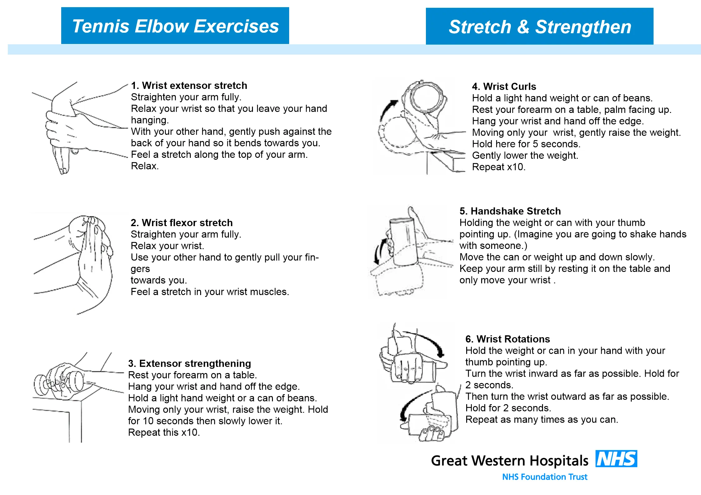
- Load management and a progressive strengthening (eccentric) exercise program is the mainstay — extensors (palm-down) for lateral, flexors (palm-up) for medial; continue exercises to prevent recurrence. Relative rest, ice, and short-term NSAIDs for symptom control; a counterforce brace may help.
- Corticosteroid injection gives short-term relief but worse medium/ long-term outcomes than exercise — reserve for severe cases.
- Surgery is rarely needed (refractory cases).
Olecranon bursitis
Swelling of the superficial bursa over the olecranon. Unlike epicondylitis above, this is a genuinely inflammatory or infective condition, not a degenerative overuse tendinopathy. Causes: trauma (acute or chronic pressure — “student’s/miner’s elbow”, e.g. truck drivers, carpet layers), rheumatoid arthritis, crystal arthropathy (gout), haemorrhage, and infection (septic bursitis). Males, age 30–60.
- Examination: localised swelling and tenderness; redness and warmth suggest an inflammatory or infective process → aspirate.
- Investigations: aspirate if red/warm/infected and send for MC&S and crystal microscopy (distinguishes septic from gout/pseudogout).
Management:
- Small, non-painful bursae need no treatment.
- Symptomatic non-infective bursitis: rest, ice, elbow pad, compression, analgesia; aspiration (therapeutic + diagnostic) for significant swelling; corticosteroid injection for refractory non-infective cases.
- Septic bursitis: aspirate and treat with antibiotics (di/flucloxacillin or cephalexin).