Plantar Fasciitis
Written 22nd February 2020 - Last Update 25th August 2026
Plantar Fasciitis
The commonest cause of inferior heel pain and the most common cause of heel pain presenting in the outpatient setting overall. Despite the “-itis” name, histology shows a degenerative, not inflammatory, process at the plantar fascia origin on the medial calcaneal tuberosity — micro-tears, collagen disarray and granulation tissue, without the inflammatory cell infiltrate the suffix implies (abductor hallucis, flexor digitorum brevis and quadratus plantae share the origin and may be involved).
- Demographics: occurs in about 10% of the general population; peak incidence 40–60 years, with 83% of patients active working adults aged 25–65. Bilateral in around a third of cases. Higher prevalence in women than men, in the 45–64 age band versus 18–44, and in those with BMI above 25 kg/m² — some series show F>M especially with obesity, consistent with this. Runner-specific prevalence is reported as high as 22%, and plantar fasciitis accounts for roughly 10% of running injuries and 11–15% of all foot symptoms presenting for care.
- Risk factors: obesity, reduced ankle dorsiflexion (tight calf/Achilles), pes planus or pes cavus, weight-bearing endurance activity (running, dancing, jumping), prolonged standing. About half of patients also have a heel spur, but the spur is a marker, not the cause. No systemic association is found in ~85% of cases; the remainder overlaps with the seronegative spondyloarthropathies.
- History:
- sharp pain under the heel, worst with the first steps in the morning (may initially walk on toes), easing with walking then building again through the day, or with prolonged standing/sitting
- recurs after sitting and towards the end of the day
- often bilateral; sometimes a history of overuse or a recent increase in weight-bearing activity.
- Examination:
- tenderness at the medial calcaneal tuberosity (~5 cm from the back of the heel); dorsiflexing the toes/foot increases the tenderness
- the windlass (Jack) test — passive dorsiflexion of the first metatarsophalangeal joint reproducing the pain — is a useful bedside confirmatory sign
- limited dorsiflexion from a tight Achilles is common; gait/heel strike otherwise normal
- tenderness over abductor hallucis suggests Baxter’s nerve (first branch of the lateral plantar nerve) entrapment.
- Differential to keep in mind: fat-pad contusion/atrophy, calcaneal stress fracture, and nerve entrapments such as tarsal tunnel syndrome — worth revisiting if the presentation is atypical or fails to settle.
- Investigations: none required for the typical presentation — clinical diagnosis. Reserve X-ray/ultrasound for atypical features or failure to improve after a reasonable trial of treatment (may show calcification, a spur, or fascial thickening); MRI if still refractory, to look for a tear, stress fracture or osteochondral defect.
- Natural history: self-limiting in most — StatPearls cites ~75% resolving spontaneously within 12 months (an older estimate often quoted as ~80%, most by 24 months); only around 5% ultimately need surgery, and even with treatment resolution can take weeks to months.
- Management (stepwise):
- Relative rest from long walks/running (return on softer surfaces), activity modification, weight loss, supportive footwear (avoid bare feet)
- Plantar-fascia-specific and Achilles/calf stretching (e.g. rolling the foot over a ball), deep friction massage, strengthening (calf raises), non-weight-bearing exercise; podiatry/physiotherapy review
- Heel pads/inner soles or prefabricated orthoses; night splints; short NSAID courses for acute pain
- Corticosteroid injection at the point of maximal tenderness. Cochrane’s review of 39 trials (2,492 adults) found only low-quality evidence of a small benefit: injected steroid modestly reduced heel pain versus placebo/no treatment in the first month, but made no difference by 1–6 months. Serious harms — fat-pad atrophy and plantar fascia rupture — were rare in the trials but likely under-reported, so can’t be ruled out as more common in practice; use sparingly and only for short-term relief, not as a default step.
- Extracorporeal shock wave therapy — second-line for >6 months of refractory pain (Cochrane rates the trial evidence for ESWT versus steroid injection as very low quality too — treat either as low-certainty, not as a clearly superior option)
- Surgery (plantar fasciotomy ± distal tarsal tunnel decompression if coexisting tibial nerve compression), open or endoscopic, reserved for persistent pain after 6–12 months of failed conservative care — complications (nerve injury, fascia rupture, arch flattening) are common and recovery protracted, and a successful outcome isn’t guaranteed.
Morton neuroma (forefoot differential)
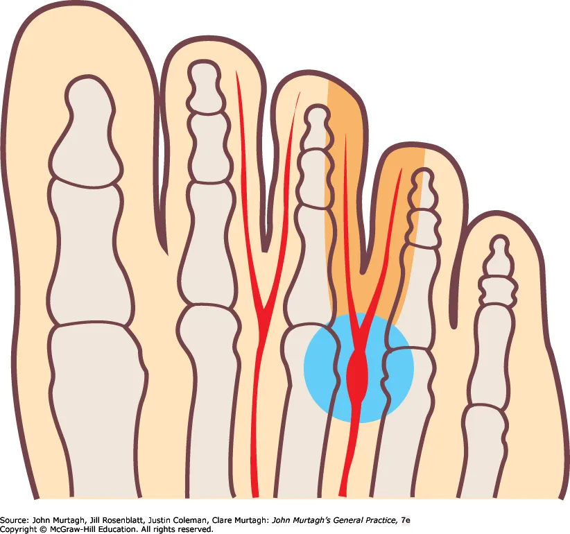
A different condition worth distinguishing from plantar fasciitis by location — not a true neuroma, but a fibrous enlargement of an interdigital nerve, most commonly between the 3rd and 4th metatarsal heads (then 2nd/3rd), rather than the heel.
- Demographics: adults <50; women 4×; bilateral in ~15%. Related to overuse and tight/inappropriate footwear.
- History: severe burning or sharp shooting pain ± paraesthesia between the affected toes; worse weight-bearing on hard surfaces and in tight shoes; relieved by removing the shoe and squeezing the forefoot.
- Examination: localised tenderness between the metatarsal heads (Mulder’s click may be felt on squeezing the forefoot).
- Investigations: clinical diagnosis; ultrasound can confirm the neuroma or show another cause (e.g. intermetatarsal bursitis).
- Management:
- Loose, low-heeled shoes + sponge-rubber metatarsal pad/orthosis to offload the nerve
- Corticosteroid injection can be considered
- Most eventually require surgical excision if conservative measures fail.