Lumbosacral Radiculopathy
Written 15th February 2020 - Last Update 25th August 2026
Lumbosacral Radiculopathy
Nerve-root (radicular/discogenic) low back pain — more complicated than non-specific LBP, but most cases resolve spontaneously: conservative management is recommended for the first 6–8 weeks, with pain usually settling gradually by 12 weeks. The commonest disc lesion is L4/5, then L5/S1 — so L5 and S1 roots account for most radiculopathy.
Presentation by nerve root
Considerable inter-patient variability and sensory overlap exist; the most reliable autonomous zones (served by a single root) are: L2–3 anterior thigh · L4 medial calf · L5 dorsum of foot · S1 sole of foot.
| Root | Pain | Power | Sensation | Reflexes |
|---|---|---|---|---|
| L1 (rare) | Inguinal region ± paraesthesia | — | Inguinal region | — |
| L2/3/4 (grouped — clinically indistinguishable) | Back → anterior thigh ± medial lower leg | Hip flexion, knee extension, hip adduction | Anterior thigh, medial lower leg | Knee ↓ |
| L5 (most common) | Back → lateral leg into foot | Foot dorsiflexion, toe extension, foot inversion/eversion | Lateral lower leg, dorsum of foot (test sharp sensation in 1st–2nd toe webspace) | Normal (internal hamstring may be ↓) |
| S1 | Back → posterior leg into foot | Plantar flexion ± hip extension, knee flexion | Posterior leg, lateral foot | Ankle ↓ |
| S2/3/4 (uncommon) | Buttock → posterior leg or perineum | Urinary/faecal incontinence, sexual dysfunction | Perineal | — |
Severity grading: 1. pure sensory/pain → 2. mild motor deficit (mild weakness or reflex loss) → 3. marked/worsening motor deficit.
Imaging
MRI is the best modality, but abnormal findings are highly prevalent in asymptomatic people — in one study of 98 asymptomatic individuals, 27% had a disc herniation and 52% a disc bulge — and do not predict future LBP. Interpret in the light of matching clinical findings.
Treatment
- Symptom relief through the acute period — disc-herniation pain usually resolves spontaneously; reassure accordingly.
- Analgesia: NSAIDs, paracetamol ± cautious short-term opioids.
- Muscle relaxants: no convincing evidence of benefit.
- Activity modification, early return to normal activity, physiotherapy; weight loss for overweight patients reduces recurrence risk.
- Epidural anaesthetic/steroid injection can be considered if not responding to conservative management.
- Degenerative arthritis/spinal-stenosis pain waxes and wanes but does worse long-term than disc herniation.
Surgical referral
- Absolute: bladder/bowel dysfunction or saddle anaesthesia (cauda equina); progressive motor disturbance (foot drop, quadriceps weakness).
- Relative: severe/prolonged pain; failure of conservative treatment; or all four of — leg pain ≥ back pain, positive straight-leg raise, no response to 4–6 weeks of conservative therapy, imaging lesion matching the symptoms.
See the low back pain (approach) hub and vertebral dysfunction with acute leg and back pain (syndrome A–D triage).
Spondylolisthesis
Forward slip of one vertebra on another (usually L5 on S1) — pain arises from stretching of the interspinous ligaments or nerve roots.
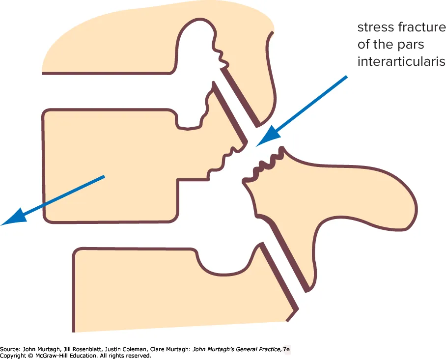
- Demographics: common (~5% of the population); many are asymptomatic and found incidentally (prevalence on imaging rises steeply with age).
- History: low back pain aggravated by prolonged standing, walking and exercise; onset generally insidious, often as concurrent disc degeneration develops.
- Examination: stiff waddling gait, increased lumbar lordosis, limited flexion; a palpable “step” may be present.
- Investigations: lateral lumbar-spine X-ray.
- Management: flexion exercise program, avoiding extension/hyperextension; analgesia and activity advice as for non-specific LBP; spinal fusion surgery for severe/intractable cases or progressive neurology.
Lumbar spondylosis (degenerative)
Degenerative osteoarthritis of the lumbar spine. May follow vertebral dysfunction or severe disc disruption: the disc desiccates and narrows, de-tensioning posterior structures; facet joints hypertrophy and degenerate, which can narrow the spinal canal and intervertebral foramina (spinal stenosis).
- Demographics: older patients.
- History: low-back stiffness is the main feature, with progressive pain on activity and improvement with rest.
- Investigations: erect plain X-ray shows the degenerative process (sclerosis, osteophytes, disc-height loss) — but degenerative findings are near universal with age and correlate poorly with symptoms.
- Management:
- basic analgesics (NSAIDs first-line as for non-specific LBP)
- exercise-based physiotherapy ± hydrotherapy
- ± TENS/acupuncture (adjuncts)
- decompressive surgery if a significant radicular/stenotic component.