Finger & Thumb Joint Injuries
Last Update 5th September 2026
Finger & Thumb Joint Injuries
For fractures of the metacarpals and phalanges see Wrist & Hand Injuries; for splint materials and technique see Fracture Immobilisation & Casting.
Assessment
Ask what direction the force came from, whether this is the dominant hand, what the patient does for work and sport, and — specifically — whether anyone reduced the finger before arrival.
Inspect for deformity, swelling, bruising and skin integrity. Dimpling or puckering of the skin over a joint suggests soft tissue interposed in it.
- Rotational deformity can’t be seen on X-rays, it is assesesed clinically. Ask for a relaxed fist with the fingertips pointing toward the scaphoid. Overlapping or “scissoring” of a digit means malrotation, and any rotational deformity needs surgical evaluation.
- Test sensation and capillary refill before injecting local anaesthetic. Compare light touch and two-point discrimination with the same digit on the other hand.
- Test the tendons individually. FDP is tested by stabilising the middle pahalx and flexing the distal phalanx at the PIP; FDS by stabilising the proximal pahalx and flexing the PIP joint. Absent or weak flexion against resistance suggests tendon damage, but pain put of proportion to the injury whuch prevents movement is also concerning.
- Test the central slip with the Elson test after any volar PIP dislocation. Flex the PIP over the edge of a table to 90° and ask the patient to extend against resistance. Weak PIP extension with a rigid DIP indicates central slip rupture and a developing boutonnière.
- Stress the joint after reduction, not before. A dorsally directed force tests the volar plate. Lateral stress is applied in both full extension and 30° of flexion; a radial force tests the ulnar collateral ligament and vice versa.
X-ray
Keep the threshold low, and take posteroanterior, oblique and true lateral views of the affected digit specifically. A history of dislocation always warrants an X-ray.
Widening of the joint space after reduction suggests interposed soft tissue, usually the volar plate. On the post-reduction lateral, the dorsal contour of the PIP joint should be a smooth “C”; a “V” shape means persistent dorsal subluxation, which leads to severe stiffness and post-traumatic osteoarthritis if left.
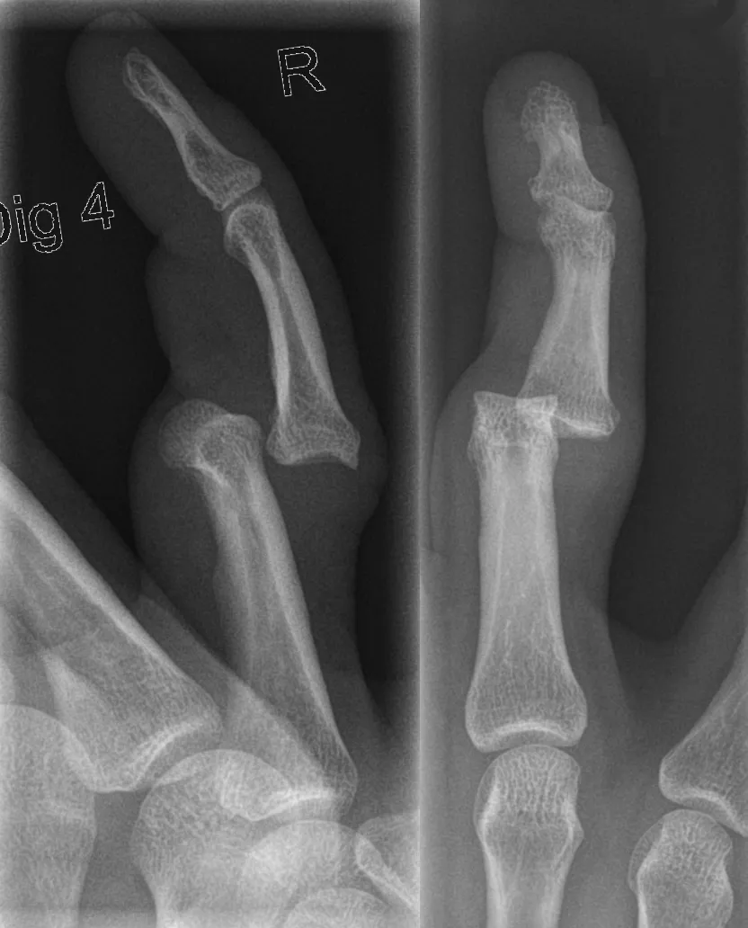
Image credit: Finger luxation D4 PIP.png — Hellerhoff, CC BY-SA 3.0, via Wikimedia Commons (https://commons.wikimedia.org/wiki/File:Finger_luxation_D4_PIP.png).

Volar plate injury and volar plate avulsion fracture
The volar plate is the fibrocartilaginous thickening on the palmar surface of the IP joints that prevents hyperextension. It tears when a ball jams the fingertip and hyperextends the PIP joint, often taking a fragment off the base of the middle phalanx with it. The injury is common, easily missed, and permanent loss of PIP joint function follows mismanagement.
Management is an extension block splint, not prolonged straight immobilisation:
- Full immobilisation for about one week only, in slight flexion or in an extension blocking splint.
- Flexion exercises start at one week, with the splint worn intermittently between sessions.
- Extension exercises start at three weeks.
- Refer if the avulsion fragment is large, or if a fragment sits within the joint space (suspect this when passive range of motion is reduced).
Refer to a hand therapist. The staged exercise-and-splint program is what determines the outcome, and hand therapists — occupational therapists or physiotherapists subspecialised in the upper limb — provide the splint as well as the program. They can be found through the Australian Hand Therapy Association (www.ahta.com.au), in private practice and in public hospitals.
PIP joint dislocation
Dorsal dislocation is the usual pattern, from longitudinal compression and hyperextension. Reduce under a digital block by applying longitudinal traction with slight extension while pushing the dorsal base of the middle phalanx volarly. Then re-examine and re-X-ray. Closed reduction and splinting are the rule unless volar plate entrapment blocks reduction, or an associated fracture renders the joint unstable.
What you find on post-reduction testing decides the treatment:
- Dislocates again with active movement — all the stabilising structures are ruptured; refer for repair.
- Hyperextends — treat as for a volar plate injury, with an extension block splint.
- Neither — splint in slight flexion for a few days, then buddy tape.
A 2014 randomised trial of buddy taping versus aluminium digital splinting for hyperextension injuries found no difference in strength, pain or function at three weeks, with earlier range of motion and less swelling in the buddy-taped group. (Paschos et al. Management of proximal interphalangeal joint hyperextension injuries: a randomized controlled trial. J Hand Surg Am. 2014 Mar;39(3):449-54)
Volar dislocations usually need referral for reduction and carry the risk of boutonnière deformity from central slip rupture. Lateral dislocations disrupt a collateral ligament and are more likely to need operative treatment; if the joint moves through a full range without subluxation after reduction, splint and reassess at two to three weeks.
Avoid over-treatment. Prolonged splinting and repeated reduction attempts at a volar PIP dislocation promote volar plate scarring and fixed flexion contracture. A fixed-flexion “pseudoboutonnière” contracture of the PIP joint can develop after a volar dislocation.
DIP joint dislocation
Usually dorsal, and often associated with a fracture or a skin injury. Reduce with longitudinal traction and a volarly directed force on the dorsal aspect of the distal phalanx with the joint flexed, then splint in slight flexion (about 10–20°) for two to three weeks if any instability persists.
An irreducible DIP dislocation means interposed volar plate, flexor digitorum profundus or terminal tendon, and needs surgery.
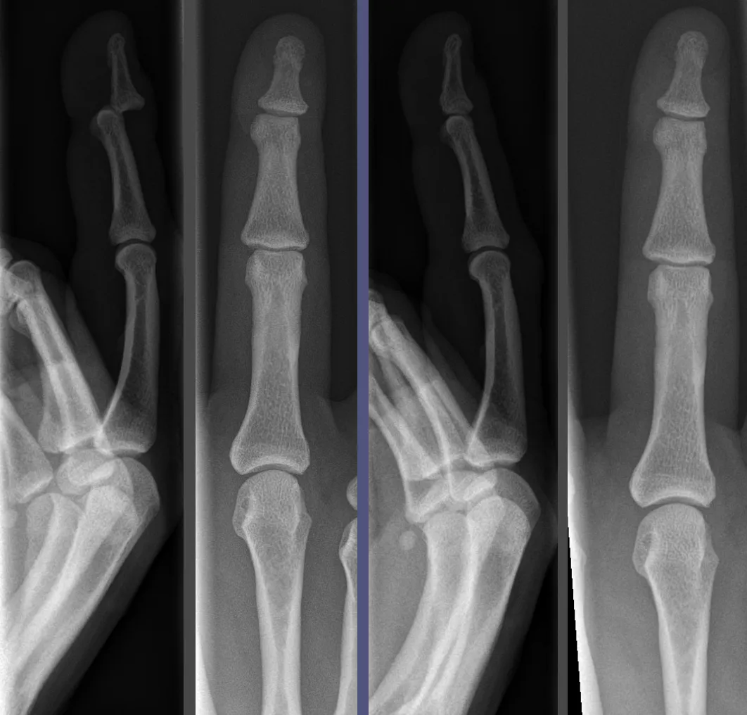
Image credit: Finger luxation pre and post.png — Hellerhoff, CC BY-SA 3.0, via Wikimedia Commons (https://commons.wikimedia.org/wiki/File:Finger_luxation_pre_and_post.png).

MCP joint dislocation
Uncommon, because the volar plate and collateral ligaments protect the joint, and usually dorsal. Reduction technique differs from the PIP joint: apply extension and axial compression over the base of the proximal phalanx to glide it back, rather than traction. Traction risks drawing the volar plate into the joint and converting a simple dislocation into a complex one.
Do not make repeated attempts. An irreducible MCP dislocation almost always means volar plate interposition and needs open reduction, and an entrapped sesamoid visible in the joint space on X-ray strongly indicates a complex dislocation. Most MCP dislocations are best referred for reduction.
Collateral ligament sprains
A sideways force at the PIP joint sprains a collateral ligament without dislocating it. Test with the joint at 30° of flexion and the MCP flexed to 90°; pain, laxity and loss of a firm end-feel indicate injury. Partial tears are buddy taped for several days followed by early active exercise.
Thumb UCL injury — skier’s and gamekeeper’s thumb
Forced abduction or hyperextension of the thumb MCP joint tears the ulnar collateral ligament. The classic mechanism is a fall onto a planted ski pole, but ball sports are the more common cause in practice. It presents with pain and swelling at the ulnar side of the thumb base and a weak, painful pinch grip. Ulnar collateral ligament injuries are around ten times more common than radial collateral ligament injuries. They account for about 86% of all athletic thumb injuries.
X-ray before stress testing — an associated avulsion fracture is a contraindication to stressing the joint.
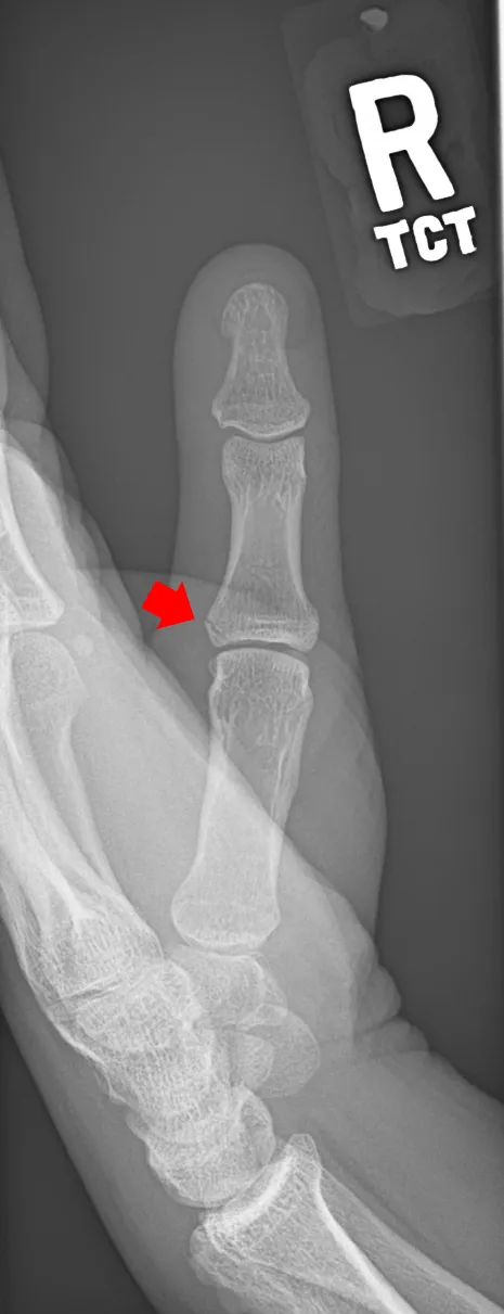
Image credit: Skiers thumb.png — Fluffy89502, CC BY-SA 4.0, via Wikimedia Commons (https://commons.wikimedia.org/wiki/File:Skiers_thumb.png).

Stress the UCL in 30° of flexion and again in full extension, comparing with the other thumb. Loss of end-feel with more than 10–20° of deviation in both positions indicates complete rupture; laxity beyond 30° is very suggestive of a tear. A palpable painful lump on the ulnar side of the joint is a Stener lesion — the torn ligament end has retracted superficial to the adductor aponeurosis, so it cannot heal in place and needs surgical repair. Where the diagnosis is equivocal on examination, MRI confirms a UCL or RCL tear with high sensitivity and specificity.
- Refer urgently to a hand surgeon for a complete rupture, a Stener lesion, or an avulsion fracture.
- Partial tears are immobilised in a thumb spica with the MCP joint in slight flexion for about six weeks, then rehabilitated with passive, active and strengthening work. Expect around six weeks before return to work or sport.
- Refer anyone with persistent pain or weakness after that period.
- Radial collateral ligament injury at the same joint is managed the same way, but a complete rupture does not automatically require surgery because no Stener lesion forms. For a complete rupture that is surgically repaired, return to sport approaches 100%.
Splinting, referral and follow-up
Splint in the safe position wherever the injury allows: at least 70° of MCP flexion, minimal PIP flexion, and 30° of wrist dorsiflexion. This holds the MCP collateral ligaments on stretch and prevents volar plate contracture. Immobilise as few joints as possible — the hand loses range quickly, so periods of full immobilisation are much shorter than for other fractures, and early movement is the goal.
Refer for same-day hand surgical assessment when there is an irreducible dislocation, an open joint injury, rotational deformity, a joint that redislocates through active range, a complete thumb UCL rupture or Stener lesion, or intra-articular involvement of more than 30% of the joint surface.
Bring the patient back with a repeat X-ray at about one week after any associated fracture, since hand fractures displace under the pull of the tendons. Warn every patient with an injured PIP joint that fusiform swelling and stiffness of that joint persist for months and are expected, so they do not interpret it as treatment failure.