Childhood Hip Disorders
Written 20th February 2020 - Last Update 14th August 2026
Childhood Hip Disorders
(Consolidates the former “Hip Paediatric — illness scripts” note, a duplicate compilation of the same five conditions.) Murtagh Ch. 65; also see hip, buttock & groin pain (approach).
Overview comparison
| DDH | Transient synovitis | Perthes | SCFE | Septic arthritis | |
|---|---|---|---|---|---|
| Age | 0–4 yrs | 4–8 yrs | 4–8 yrs | 10–15 yrs | All ages |
| Limp | + | + | + | + | Refuses to walk |
| Pain | No | + | + | + | +++ |
| ROM | Abduction limited | Limited all, esp. internal rotation & abduction | Limited abduction & internal rotation | Limited all, esp. internal rotation | Extremely limited |
| X-ray | Negative early | Normal | Subchondral fracture, dense head, pebble-stone epiphysis | Slip in epiphysis | Normal |
| USS | Up to 6 months | — | — | — | Can show effusion |
Developmental dysplasia of the hip (DDH)
- Demographics: females 6:1; the classic 5 F’s — Female, first-born, family history, foot abnormalities, “funny” deliveries (breech/transverse, oligohydramnios, C-section).
- History: may present with a shortened leg from birth to age 4; painless limp once walking — should ideally be detected before this to allow early correction.
- Examination: Barlow and Ortolani tests (best before 3 months, usually negative after); asymmetric gluteal skin folds.
- Investigations: ultrasound is gold standard — do it for breech delivery or any of the 5 F’s risk factors; USS up to 6 months (not in the first 6 weeks; unreliable 4–6 months — consider X-ray in that window). X-ray has no diagnostic value in the neonatal period but is useful after 6 months.
- Management:
- Positive Barlow/Ortolani → urgent orthopaedic referral + organise USS
- 0–6 months: Pavlik harness or abduction splint
- 3–18 months: closed or open reduction + pelvic spica cast
- >18 months: open reduction ± osteotomy
- Despite treatment, some progress to acetabular dysplasia and premature OA needing joint replacement — consider follow-up X-ray in the teenage years for anyone with a DDH history.
Perthes disease (idiopathic avascular necrosis of the femoral head)
A diagnosis of exclusion: over months to years the femoral head undergoes necrosis, fragmentation, then healing with a residual flat spot.
- Demographics: 4–8 yrs; boys 4×; ~5–15 per 100,000 births.
- History: hip ache with an atraumatic limp.
- Examination: antalgic gait; restricted internal rotation and abduction.
- Investigations: X-ray (may be normal early — a crescent sign heralds the fragmentation phase, with later reduced femoral head size); if normal but concern remains, MRI is MBS-covered.
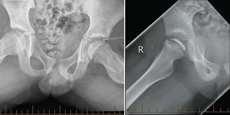
- Management: “containment” — keep the femoral head within the acetabulum so it heals spherical. Controversial and orthopaedics-led: non-weight-bearing, ROM exercises, observation, or osteotomy depending on severity.
Slipped capital femoral epiphysis (SCFE)
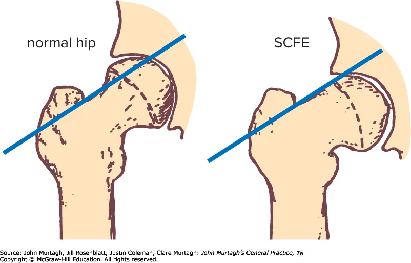
The femoral head slips posteriorly and inferiorly relative to the neck (the head stays in the acetabulum; the neck displaces) through the growth plate. Mostly idiopathic; consider secondary causes that weaken bone — trauma, obesity, endocrine disease, renal failure, osteodystrophy, radiation.
- Demographics: 8–15 yrs (peak incidence, earlier in girls than boys); obesity (~80% of cases) and growth spurts are the main associations; males > females. Bilateral in up to 40% — of these, most slip sequentially rather than simultaneously (usually within 18 months of the first side), so a normal contralateral hip at diagnosis doesn’t rule out a later slip on that side; compare both hips carefully on imaging.
- History: hip/groin/thigh/knee pain with a limp, worse with activity — 90% present as “stable” (progressive pain/limp over weeks to months, still able to weight-bear ± crutches); the ~10% “unstable” presentation (sudden inability to weight-bear at all, often after minor trauma) is a much more urgent problem with a higher AVN risk.
- Examination: leg shortened and externally rotated; restricted internal rotation is usually the first movement lost. Obligatory external rotation on hip flexion (Drehmann sign) — flexing the hip passively causes it to rotate externally rather than staying neutral — is a useful supporting sign.
- Investigations: AP + frog-leg lateral X-ray of both hips (compare sides — early slippage can be subtle and near-symmetrical if bilateral). Look for the line of Klein (drawn along the superior femoral neck) — normally intersects a small portion of the epiphysis; in SCFE the line passes above the epiphysis without intersecting it. The classic description is the epiphysis looking like “ice cream falling off the cone. “If concern remains despite a normal/equivocal X-ray, MRI or ultrasound (up to 95% sensitive) can help.
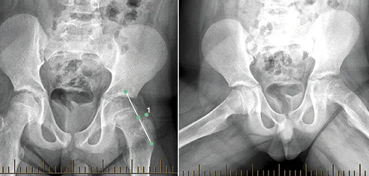
- Management: cease weight-bearing immediately (crutches/wheelchair, even in a “stable” slip that can technically still bear weight) and refer to orthopaedics urgently (likely via ED) — a stable slip needs fixation within days; an unstable slip (can’t weight-bear at all) needs emergency referral, with a significantly higher risk of avascular necrosis. Definitive treatment is single-screw in-situ fixation. Missed SCFE → avascular necrosis (10–15% of cases, higher with instability, greater slippage or repeated reduction attempts) and chondrolysis → early osteoarthritis.
Transient synovitis
Self-limiting synovial inflammation, usually post-viral.
- Demographics: 3–8 yrs (peak 6).
- History: sudden unilateral hip pain and limp; often recent trauma or URTI/viral illness (within 2 weeks).
- Examination: painful limitation of all movement, especially abduction/ internal rotation.
- Investigations: ESR/CRP may be mildly elevated; usually afebrile; USS shows joint fluid; no organism on aspiration. If afebrile with normal CRP, septic arthritis is unlikely — but let orthopaedics make that call.
- Management: refer early to exclude septic arthritis (may need image-guided ± anaesthetic aspiration); usually settles spontaneously by 7 days; X-ray at 4–6 months to exclude evolving Perthes disease.
Septic arthritis
Suspect in any child with an acutely painful or irritable hip — the child may not look obviously unwell but will refuse to walk, with pain on all movements. A negative aspirate does not exclude septic arthritis — if fever persists with a negative aspirate, proceed to arthrotomy. Can only be excluded once X-ray, USS, bloods and bone scan are all negative, at which point a diagnosis of irritable hip syndrome can be made.