Ankle tendon disorders
Written 22nd February 2020 - Last Update 20th August 2026
Ankle tendon disorders
Tenosynovitis around the ankle follows repetitive overuse, trauma (e.g. an ankle sprain) or unaccustomed stress — friction where the tendons angulate at the ankle. Most commonly tibialis posterior (medial) or the peroneal tendons (lateral); occasionally tibialis anterior and extensor digitorum longus. Non-infective tenosynovitis more broadly is overuse, autoimmune (rheumatoid arthritis and other inflammatory arthritides) or idiopathic — take a history of fluoroquinolone use and prior corticosteroid injection, both independently associated with tendon disease and rupture.
Presentation: pain, swelling and restricted movement, with tenderness where the tendon bulges from behind/below the malleolus; a history of snapping or popping at the malleolus suggests subluxation rather than simple tendinopathy. Ultrasound is a cheap, radiation-free first investigation and can also guide injection; MRI confirms if needed, with higher sensitivity for partial tears. Complications: tenovaginitis, weakness, ganglion formation, subluxation/dislocation, rupture.
General treatment: partial immobilisation (rarely a cast) or an orthotic arch support; a carefully directed corticosteroid injection into the tendon sheath can be very effective (avoid intratendinous injection) — sheath injection can also help confirm the diagnosis if the response is dramatic. Add NSAIDs and activity modification (footwear change, a lateral heel wedge for peroneal-side pain) as first-line adjuncts; physiotherapy guides the rehabilitation phase. Give a non-operative trial of 4–6 months before considering surgery, which is reserved for those who fail conservative care or have a structural lesion (tear, subluxation, retinaculum injury) unlikely to settle on its own.
Peroneal tendinopathy
Along the tendon course from behind the lateral malleolus to the outer foot; common in athletes and ballet dancers. The peroneus brevis and peroneus longus share a common synovial sheath behind the fibula before separating, running through a tunnel bordered by the superior peroneal retinaculum, the fibula’s retromalleolar groove, and the calcaneofibular ligament — pain on palpation, passive inversion, or resisted eversion. Chronic lateral ankle instability is itself a recognised cause of peroneal tendon injury (repeated subluxation over the fibula causing longitudinal tears), so a peroneal problem after a “sprain that never settled” should prompt a specific look for tendon pathology, not just persistent ligament laxity. An anatomically shallow or flattened retromalleolar groove, hindfoot varus, or a low-lying peroneus brevis muscle belly all predispose to subluxation and tearing.
Peroneal tendon tears are most often longitudinal split tears from chronic subluxation over the fibula (typically peroneus brevis, ~88% of tears) rather than a single traumatic transverse tear; consider in a patient with constant swelling, pain, or a subjective sense of ankle instability or weakness rather than a discrete pop. Weight-bearing ankle and foot X-rays (AP, mortise, lateral, plus a Harris heel view) look for an avulsion at the base of the fifth metatarsal or distal fibular groove, an os peroneum, or groove flattening. Ultrasound is sensitive (~100%) and specific (~85%) for tears and doubles as a dynamic test for subluxation; MRI is slightly less sensitive (~83%) but adds detail on bony deformity and oedema. Rule out ankle sprain, calcaneal fracture, fractured os peroneum and rheumatoid arthritis as differentials; a varus heel with peroneal disease raises the possibility of underlying Charcot-Marie-Tooth disease.
Peroneal tendon dislocation/subluxation — classically forceful dorsiflexion with reflex peroneal contraction against a taut superior peroneal retinaculum; an audible (often painless) snap, with the tendons visibly/palpably slipping anterior over the fibula on active eversion. Confirm on exam with the patient prone, knee flexed to 90°, testing for subluxation on resisted eversion. Acute instability follows a torn superior peroneal retinaculum or a retromalleolar groove avulsion; chronic subluxation instead reflects groove flattening and retinacular laxity, and carries a real risk of being mistaken for (or coexisting with) a simple ankle sprain if not specifically examined for. Requires surgical repair — options include retinaculum repair/reconstruction, fixation of an avulsion fracture, fibular groove deepening for a shallow groove, correction of hindfoot varus, or tendon rerouting/bone-block procedures for recurrent instability — outcomes are good, with return to sport reported in the large majority of surgically treated patients.
Tibialis posterior tendinopathy (posterior tibial tendon dysfunction)
Common in middle-aged women, ballet dancers, and pes planus with valgus deformity — affecting an estimated 3% or more of women over 40, and associated with obesity, diabetes, hypertension and previous corticosteroid injection near the tendon. The tendon (foot invertor) inserts on the navicular tuberosity, and runs through a relatively hypovascular “watershed” zone just posterior to the medial malleolus that predisposes it to degeneration and poor healing after injury.
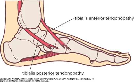
- Pain/weakness in the medial ankle and foot, worse standing and walking; standing on toes painful and difficult
- Tenderness anterior/inferior to the medial malleolus; pain on passive eversion stretch and on resisted inversion
- May cause tarsal tunnel syndrome
- Diagnosis: ultrasound; MRI is gold standard for tears — a weight-bearing radiograph (AP, lateral, hindfoot views) is the mainstay for staging the resulting deformity, and ultrasound of the tendon itself correlates with MRI in roughly 87–94% of patients
- Treatment: conservative, with good outcome over 12–24 months — bilateral semi-rigid orthotic arch support, physiotherapy-guided exercises, massage; ultrasound-guided sheath steroid injection best avoided; surgical opinion if conservative care fails
Tibialis posterior tendon dislocation — forceful dorsiflexion+inversion; painful, can’t weight-bear, tendon may be visible over the medial malleolus; immediate surgical repair. This is a rare injury, easily missed and misdiagnosed as a simple ankle sprain, so keep it in mind for a “sprain” with visible/palpable anterior displacement of the tendon at the medial malleolus rather than lateral tenderness alone.
Tibialis posterior tendon rupture — relatively common and misdiagnosed, especially in middle-aged women, and predisposed to by the same hypovascular watershed zone, prior corticosteroid injection, and chronic tendinopathy that underlie the more gradual dysfunction above. Often little discomfort at the moment of rupture; presents later with a sudden “abnormal” flat foot (collapsed longitudinal arch) and gross eversion.
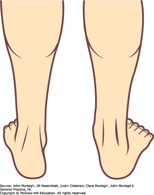
- “Too many toes” test — viewed from ~3 m behind, more toes are visible on the affected side; the single heel raise test is also diagnostic (can’t raise, or no heel inversion).
- Ultrasound is the most useful investigation. Minor cases: orthotics; severe: surgical correction (responds well).