Leishmaniasis
Written 15th August 2020 - Last Update 30th August 2026
Leishmaniasis
A protozoan (Leishmania spp.) transmitted by sandfly bite. Not endemic to Australia — seen here only in travellers/migrants from endemic regions (Asia, Middle East, North Africa, the Mediterranean, and South/Central America; 1.5-2 million new cases/year worldwide). Reservoirs vary by species/region — dogs, rodents, other animals; humans for L. donovani on the Indian subcontinent. Three clinical forms: cutaneous, mucosal, visceral.
Who to suspect it in: a returned traveller or migrant with a chronic non-healing skin ulcer, or unexplained fever with hepatosplenomegaly/pancytopenia, after time spent in an endemic area — ask specifically about rural/outdoor exposure and sandfly bites.
Clinical forms
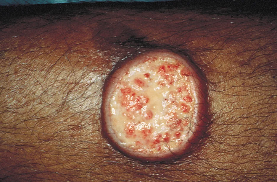
- Cutaneous (the most common form) — incubation 2-4 weeks: an asymptomatic papule at the bite site enlarges into a well-circumscribed, painless ulcer with a raised border. Heals spontaneously over 2-5 years (species-dependent), leaving a depressed scar. Diffuse cutaneous disease can involve the whole skin surface and progress to leonine facies.
- Mucosal — from hematogenous/lymphatic spread, usually after a cutaneous ulcer has healed; onset within 2 years but can be delayed decades. Progressive naso-oropharyngeal ulceration (nose/mouth mucosa; vocal cords and trachea can be involved) — can be severe and life-threatening, though bone is spared.
- Visceral (kala-azar) — gradual fever, hepatosplenomegaly, pancytopenia, hypergammaglobulinaemia; fatal if untreated. Reactivates with immunosuppression (HIV/AIDS) — mortality in HIV-positive patients is far higher than HIV-negative (33.6% vs 3.6% in one Ethiopian cohort).
Diagnosis
Histopathology/culture of tissue is the gold standard — Giemsa-stained smear or impression smear from the ulcer margin (cutaneous/mucosal), or bone marrow/spleen/ lymph-node aspirate (visceral). PCR is sensitive and specific and increasingly the test of choice. Serology has poor sensitivity in leishmaniasis (weak humoral response) so is used more for visceral disease than cutaneous.
Management
- Refer to infectious diseases/tropical medicine — species identification (guides drug choice and resistance risk) and treatment are specialist-led.
- Localised cutaneous disease often resolves spontaneously; first-line is intralesional pentavalent antimonials (sodium stibogluconate, meglumine antimoniate), or heat/cryotherapy for fewer than 5 lesions. Alternatives: systemic miltefosine, amphotericin B, pentamidine, paromomycin.
- Mucosal or complex cutaneous disease: systemic therapy — liposomal amphotericin B achieves ≥90% cure rates; pentavalent antimonials are WHO first-line but less effective than amphotericin.
- Visceral disease requires treatment; regimen depends on region because resistance patterns vary widely (e.g. sodium stibogluconate works well in East Africa but has high failure rates in India). Liposomal amphotericin B and oral miltefosine are the most widely used agents; miltefosine is the only oral option for visceral disease.
- Prevention — sandfly avoidance (fine-mesh permethrin-treated bed nets — sandflies are smaller than mosquitoes and penetrate standard nets), DEET, and reservoir/vector control (dog vaccination/insecticide collars in endemic areas).