Neutrophilia & Lymphocytosis
Written 22nd July 2020 - Last Update 30th August 2026
Presentation: Neutrophilia/Lymphocytosis
Total white blood cell count alone can be misleading — always read the differential and, if it is abnormal, a manual film.
In adults the normal WBC is roughly 4.0–11.0×10⁹/L; a count above 11.0×10⁹/L is leukocytosis, and above 100×10⁹/L is hyperleukocytosis. Leukocytosis is classified by the predominant cell type; neutrophilia is the commonest form.
Normal neutrophil range roughly 2.0–8.0×10⁹/L; normal lymphocyte range roughly 1.0–4.0×10⁹/L (lymphocytes are about 20–40% of the WBC).
Neutrophilia
Neutrophil facts: neutrophils are the first cells recruited to a site of infection or injury and are the hallmark of acute inflammation.
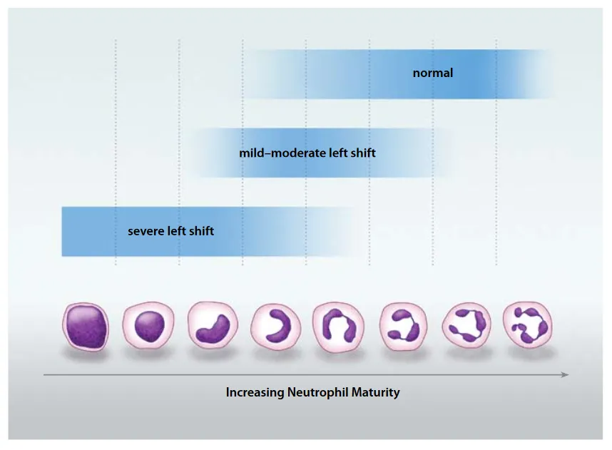
- In infection, more immature forms (bands, metamyelocytes) are released early — a “left shift.”
- Severe infection can show toxic granulation and Döhle bodies on the film.
- Neutrophils rise with almost any acute stress — vigorous exercise, temperature change, seizures, surgery, smoking — because stress hormones (catecholamines, glucocorticoids) mobilise them from the marginated and marrow pools (demargination).
Causes:
- Reactive (secondary) — the great majority: infection and inflammation (bacterial more than viral), tissue necrosis, chronic inflammatory disease (rheumatoid arthritis, adult Still disease, vasculitis, inflammatory bowel disease, chronic hepatitis), solid-tumour malignancy (paraneoplastic or marrow metastasis), pregnancy, obesity, asplenia/hyposplenism, haemolysis.
- Drugs: glucocorticoids, G-CSF/GM-CSF, all-trans retinoic acid, lithium, catecholamines (adrenaline).
- Stress/physiological: heavy exercise, emotional or physical stress, cigarette smoking.
- Primary (clonal): myeloproliferative neoplasms — chronic myeloid leukaemia, polycythaemia vera, essential thrombocythaemia, chronic neutrophilic leukaemia; chronic idiopathic (constitutional) neutrophilia, where the count sits just above the reference range with no pathology (about 2.5% of healthy people meet the statistical definition — a family member’s count can help).
- Factitious: platelet clumping or cryoglobulin precipitation in a badly anticoagulated sample — a repeat sample and film resolve it.
Leukemoid reaction: a WBC over 50×10⁹/L driven by a reactive cause (Clostridioides difficile, tuberculosis, pertussis, some non-haematological cancers, drugs such as ATRA). It is distinguished from leukaemia by a predominance of mature neutrophils rather than blasts, and it resolves when the cause is treated.
Red flags: person particularly unwell; severity of neutrophilia; rate of change; presence of a left shift; constitutional symptoms (weight loss, night sweats, fever), lymphadenopathy or hepatosplenomegaly.
When to think about CML: a persistently rising neutrophil count can be the presenting sign of chronic myeloid leukaemia.
Characteristic changes are a moderate neutrophil increase (over 50×10⁹/L) with a left shift and prominence of myelocytes on the film. Chronic mild neutrophilia without a left shift is very unlikely to be chronic myeloid leukaemia.
If a myeloproliferative neoplasm is suspected, the film plus molecular testing (BCR-ABL1 for CML) and flow cytometry sort it out, with a bone marrow biopsy where needed.
What to do next: for essentially well people with an incidental FBC finding — repeat in 1–2 months, continue following 3-monthly for 12 months then occasionally thereafter, and stop once the count has normalised.
If a cause is suspected, targeted tests help separate reactive from clonal: inflammatory markers (ESR, CRP, LDH, ferritin), liver and renal function, and cultures for suspected infection.
Lymphocytes
Lymphocytosis
Lymphocytosis is an absolute lymphocyte count above 4.0×10⁹/L in adults (relative lymphocytosis is a lymphocyte fraction over 40% with a normal absolute WBC). Different subsets (T, B or NK cells) rise depending on cause.
Reactive lymphocytes are pleomorphic (varied); a monomorphic population suggests a clonal disorder.
Causes:
- Acute infection (usually self-limiting): Epstein–Barr virus (infectious mononucleosis — lymphocytosis in up to two-thirds of cases), cytomegalovirus, acute (primary) HIV, viral hepatitis, influenza, mumps, measles, rubella; Bordetella pertussis and Bartonella henselae (cat-scratch disease) are bacterial exceptions to the usual neutrophil response; toxoplasmosis, tuberculosis.
- Stress: transient lymphocytosis after a seizure, trauma, cardiac event or adrenaline — it precedes the later neutrophilia.
- Other reactive: smoking (persistent polyclonal B-cell lymphocytosis in young women who smoke), hyposplenism/post-splenectomy (a stable long-term rise), autoimmune thyroiditis, drug reactions (DRESS with allopurinol, carbamazepine, vancomycin, sulfonamides).
- Lymphoproliferative disorders: chronic lymphocytic leukaemia (the commonest adult leukaemia, median age ~70, needs a sustained clonal lymphocyte count ≥ 5.0×10⁹/L), monoclonal B lymphocytosis (< 5.0×10⁹/L clonal B cells, no other features — a CLL precursor state, prevalent in 5–9% of people over 60), non-Hodgkin lymphomas (mantle cell, marginal zone, follicular, hairy cell), large granular lymphocyte leukaemia, and adult T-cell leukaemia/lymphoma (HTLV-1).
Consider CLL and other lymphoproliferative disorders in persistent lymphocytosis — rare below age 40, normally an incidental finding, may be associated with lymphadenopathy, hepatomegaly and splenomegaly.
Follow-up: if essentially well with no associated features, check in 1–2 months. If mild symptoms of a transient condition (e.g. infection), recheck once well.
Anaemia or thrombocytopenia alongside the lymphocytosis, or blasts on the film, raises concern for a clonal disorder and dictates urgency. Persistently elevated lymphocytes with lymphadenopathy/hepatomegaly/ splenomegaly should be referred for haematology assessment.
A note on lymphocyte surface markers (flow cytometry): a costly test that should not be ordered routinely.
Reasonable indications include a persistent unexplained lymphocytosis beyond one month, a very high count (an absolute lymphocyte count over ~30×10⁹/L is one cited threshold), abnormal lymphocyte morphology or blasts on the film, other abnormal blood-count parameters (anaemia, thrombocytopenia), or lymphadenopathy/hepatosplenomegaly once reactive causes are excluded.
Flow cytometry establishes clonality and the immunophenotype (e.g. CLL cells are CD5+, CD23+, CD20-dim, CD200+); FISH and mutation analysis then refine the diagnosis and risk.
Infectious mononucleosis management is supportive (analgesia/NSAIDs); because most patients have splenomegaly, contact sport should be avoided in the early acute phase to prevent splenic rupture.